
|
Fig. 2
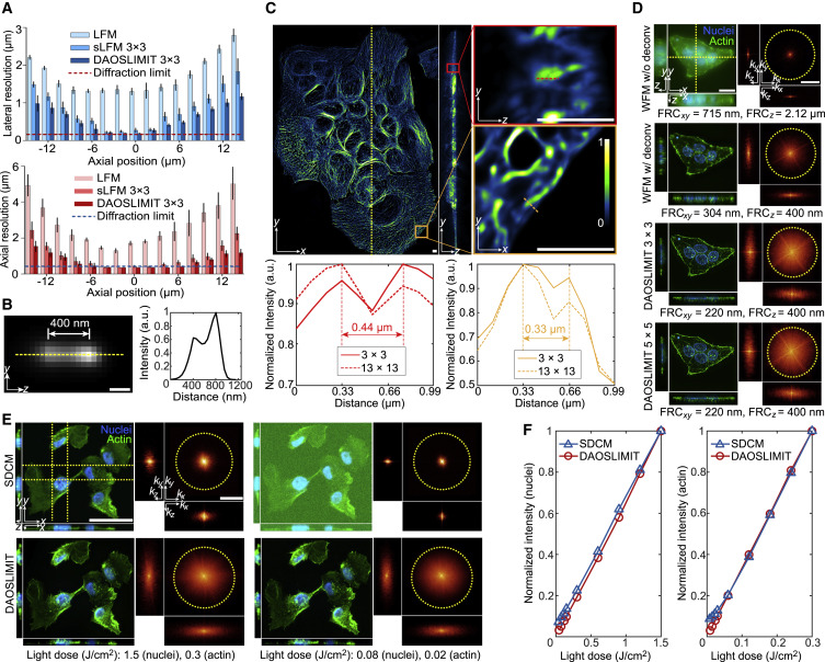
Figure 2. Resolution and SNR characterization of DAOSLIMIT (A) Boxplots of the lateral and axial resolutions of LFM, sLFM (DAO off), and DAOSLIMIT at different axial positions with 63×/1.4 NA oil-immersion objective. The resolution is estimated by the FWHMs of the intensity profiles with a Gaussian fit for 100-nm-diameter beads distributed in 1% agarose (n = 300 beads, 20 beads per plane). Error bars represent SD. (B) MIP of two beads in yz plane obtained by DAOSLIMIT at the focal plane with a cross-section profile. We imaged the same bead at two axial planes separated by 400 nm and added the images to create virtually spaced beads. (C) Orthogonal MIPs from 12-μm-thick slabs of MCF10A cells labeled with microtubules imaged by DAOSLIMIT (3 × 3 scanning), with normalized profiles of the marked lines. (D) Orthogonal MIPs from 16-μm-thick slabs of HeLa cells labeled with actin (green) and nuclei (blue), which were obtained by WFM (90 axial slices at 200-nm steps), WFM with 3D deconvolution, and DAOSLIMIT with 3 × 3 and 5 × 5 scannings. The Fourier transforms of the MIPs are shown with estimated resolutions by FRC. Throughout the figure, the yellow dashed circles correspond to the Abbe diffraction limit of the objective. (E) Orthogonal MIPs and corresponding Fourier transforms of the MCF10A cells with actin (green) and nuclei (blue) labeling imaged by SDCM (1.3 NA) and DAOSLIMIT (1.4 NA) under different light doses used to obtain the volume marked at the bottom. The yellow dashed lines indicate the regions for yz and xz projections. (F) The normalized average intensities of the two channels were plotted against different light doses. Scale bars: 200 nm (B); 4 μm (C); 20 μm, 4 μm−1 (D); and 50 μm, 4 μm−1 (E). See also Figures S3 and S4.
Reprinted from Cell, 184, Wu, J., Lu, Z., Jiang, D., Guo, Y., Qiao, H., Zhang, Y., Zhu, T., Cai, Y., Zhang, X., Zhanghao, K., Xie, H., Yan, T., Zhang, G., Li, X., Jiang, Z., Lin, X., Fang, L., Zhou, B., Xi, P., Fan, J., Yu, L., Dai, Q., Iterative tomography with digital adaptive optics permits hour-long intravital observation of 3D subcellular dynamics at millisecond scale, 3318-3332.e17, Copyright (2021) with permission from Elsevier. Full text @ Cell