
|
Fig. 1
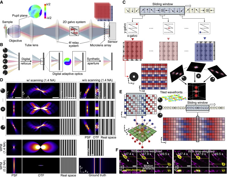
Figure 1. Principle of DAOSLIMIT (A) Schematic of the sLFM system for high-resolution spatial-angular measurements. (B) Illustrations of DAO. Different angular components can be manipulated in postprocessing for 3D reconstruction and aberration corrections. (C) Illustrations of the periodic scanning process and pixel realignment. Different colors correspond to specific galvo positions (indicated by the arrows) with a period of 9 for 3 × 3 scanning. The sensor pixels with the same relative position to the center of each microlens belong to the same angle. Images at different scanning positions can be realigned together to increase spatial sampling for 3D reconstruction with tiled aberration corrections digitally. (D) Comparisons between the PSFs, OTFs, and simulated imaging results of LFM, sLFM, and WFM with a 63×/1.4 NA oil-immersion objective and WFM with a 63×/0.2 NA air objective at a center wavelength of 525 nm. The simulated sample is a high-frequency sinusoidal pattern with a period of 1.3 μm, as shown in the bottom right corner. To show the highest contrast, the sample is placed at the native focal plane for WFM and 1 μm away from the focal plane for LFM and sLFM. (E) Illustrations of the time-weighted algorithm. High-resolution time-lapse data can be obtained by applying inverse distance weighting in the xyt domain with a sliding window. (F) Comparisons between the reconstructed results (MIP) of DAOSLIMIT with and without the time-weighted algorithm. Scale bars: 5 μm, 2 μm−1 (D) and 10 μm (F). See also Figures S1 and S2 and Video S1.
Reprinted from Cell, 184, Wu, J., Lu, Z., Jiang, D., Guo, Y., Qiao, H., Zhang, Y., Zhu, T., Cai, Y., Zhang, X., Zhanghao, K., Xie, H., Yan, T., Zhang, G., Li, X., Jiang, Z., Lin, X., Fang, L., Zhou, B., Xi, P., Fan, J., Yu, L., Dai, Q., Iterative tomography with digital adaptive optics permits hour-long intravital observation of 3D subcellular dynamics at millisecond scale, 3318-3332.e17, Copyright (2021) with permission from Elsevier. Full text @ Cell