
Image
|
Figure Caption
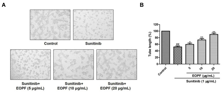
Fig. 7
EOPF promoted tube formation of HUVECs. (A) A microscope was used to photograph tubular structures (100 × magnification). (B) EOPF treatment at 5, 10, and 20 µg/mL significant boosted tube formation in sunitinib-injured HUVECs. Results are presented as the mean ± SD for three individual experiments. ##P < 0.01 vs untreated control, **P < 0.01 vs sunitinib, *P < 0.05 vs sunitinib.
Acknowledgments
This image is the copyrighted work of the attributed author or publisher, and
ZFIN has permission only to display this image to its users.
Additional permissions should be obtained from the applicable author or publisher of the image.
Full text @ Drug Des Devel Ther