
Image
|
Figure Caption
Fig. 4
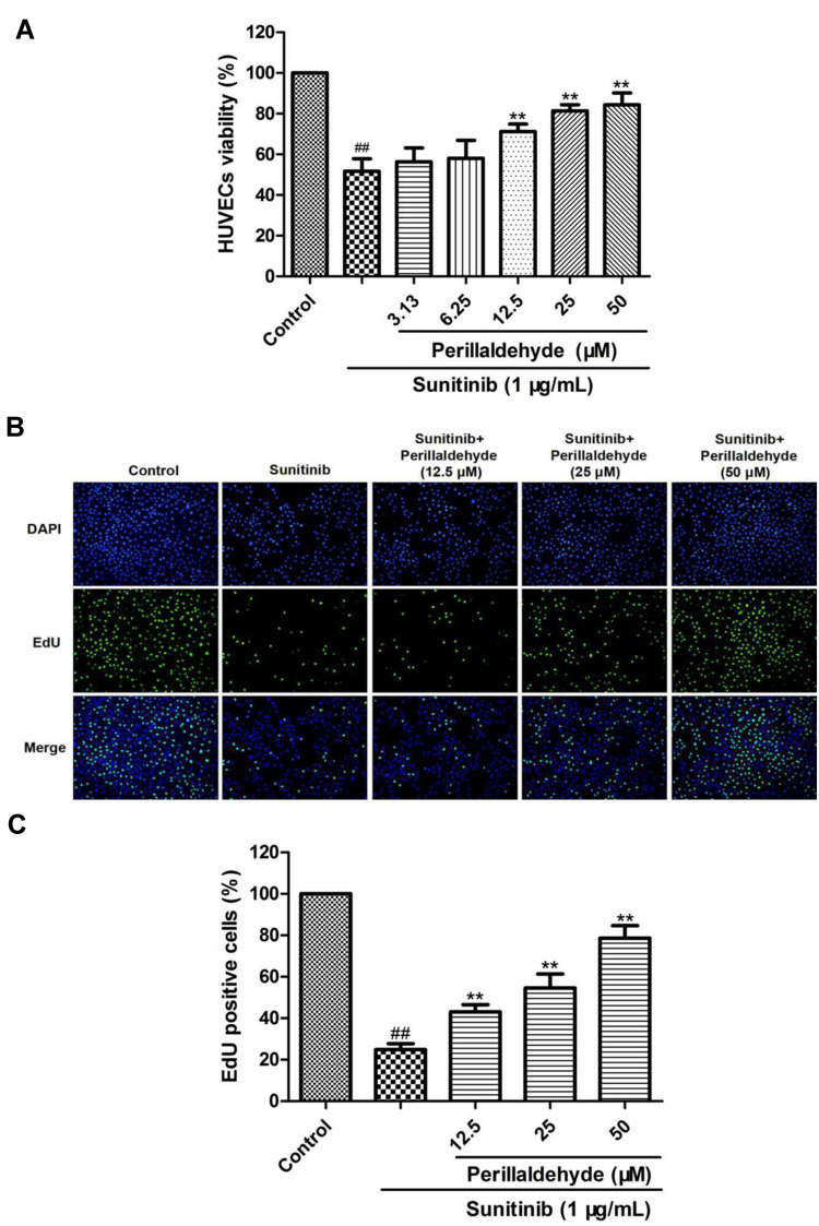
Perillaldehyde promoted proliferation of HUVECs. (A) An MTT assay was carried out to measure HUVECs viability. Perillaldehyde at 12.5, 25, and 50 µM signally increased viability of sunitinib-treated HUVECs. (B) Cells of DAPI and EdU staining were captured. (C) Perillaldehyde at 12.5, 25, and 50 µM observably promoted proliferation of sunitinib-injured HUVECs. Results are presented as the mean ± SD for three individual experiments. ##P < 0.01 vs untreated control, **P < 0.01 vs sunitinib.
Acknowledgments
This image is the copyrighted work of the attributed author or publisher, and
ZFIN has permission only to display this image to its users.
Additional permissions should be obtained from the applicable author or publisher of the image.
Full text @ Drug Des Devel Ther