
Image
|
Figure Caption
Fig. 4
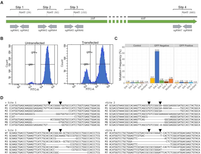
GFP Knockout Screen. (A) Genomic DNA was isolated from HeLa cells stably expressing GFP-LC3. The four HpaII sites within the GFP ORF and the predicted sgRNAs are labeled. (B) FACS sorting of transfected and untransfected cells. GFP fluorescence is on the x-axis and counts are on the y-axis. (C) Mutation frequency at each of the HpaII sites based on high-throughput sequencing of the GFP locus. (D) The most common variants for each of the four HpaII sites.
Acknowledgments
This image is the copyrighted work of the attributed author or publisher, and
ZFIN has permission only to display this image to its users.
Additional permissions should be obtained from the applicable author or publisher of the image.
Full text @ Nucleic Acids Res.