IMAGE

FIGURE 8

ID
ZDB-IMAGE-230115-13
Source
Figures for Zhu et al., 2023
Image
Figure Caption

FIGURE 8

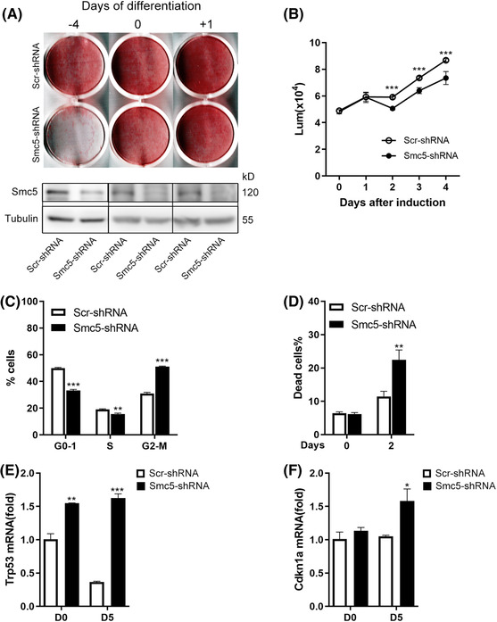
Smc5 is essential for mitotic clonal expansion during adipogenesis. (A) Representative stained plates (top) and Western blots (bottom) of 3T3‐L1 cells infected with Smc5 or scrambled shRNA at different time points, followed by induction of adipogenesis. (B) Relative proliferation of 3T3‐L1 white preadipocyte cells before and after differentiation was assessed by CellTiter‐Glo assay, n = 5. (C) Cell cycle profile by fluorescence‐activated cell sorting (FACS) at D2 of adipogenesis. (D) Quantification of dead cells (7‐AAD) by FACS at D0 and D2 of adipogenesis. (E and F) Quantitative real‐time PCR (qPCR) of Trp53 (p53) and Cdkn1a (p21) expression at D0 and D5 of adipogenesis. Rplp0 (36B4) was chosen as the internal reference gene. The data are presented as the mean ± s.d. in (B–D) and mean ± s.e.m. in (E and F). For parts (B–F), the p values were calculated using a two‐sided Student's t‐test. *p < .05, **p < .01 and ***p < .001

Acknowledgments
This image is the copyrighted work of the attributed author or publisher, and ZFIN has permission only to display this image to its users. Additional permissions should be obtained from the applicable author or publisher of the image. Full text @ Clin Transl Med