
|
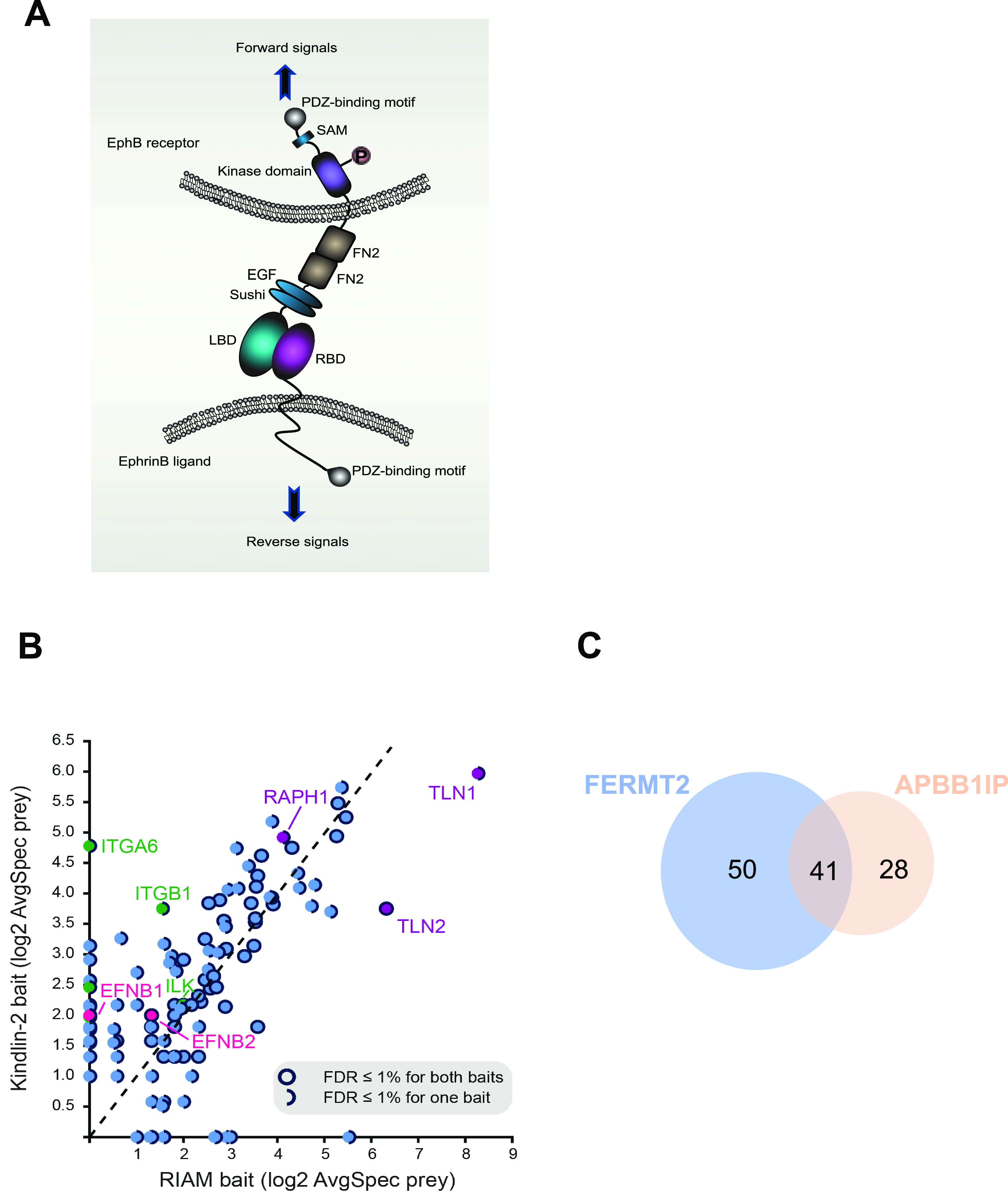
Figure 1.
(A) Domain structure and bi-directional signaling of ephrinB and EphB. EphB receptors and ephrinBs located at the cell membrane of opposing cells interact in trans and activate short-distance bi-directional signaling. LBD, ligand-binding domain; RBD, receptor-binding domain; FN, fibronectin domain; EGF, epidermal growth factor–like domain; SAM, sterile alpha motif. Orange circle indicates tyrosine phosphorylation. (B) Scatterplot comparing the averaged spectra (log2-transformed) for high-confidence preys identified from kindlin2 with RIAM baits; notable preys are indicated and filled in different colors, with details in the legend inset. (C) Venn diagram showing the number of overlapped preys discovered in the proximity interactomes of kindlin2 and RIAM baits. All high-confidence interactions (AvgP ≥ 0.95) for each bait were considered. See Fig S1 for details.