
Figure 4
- ID
- ZDB-IMAGE-221226-331
- Genes
- Antibodies
- Publication
- Weeks et al., 2022 - Embryonic alcohol exposure disrupts the ubiquitin-proteasome system
- All Figures
- Figures for Weeks et al., 2022
|
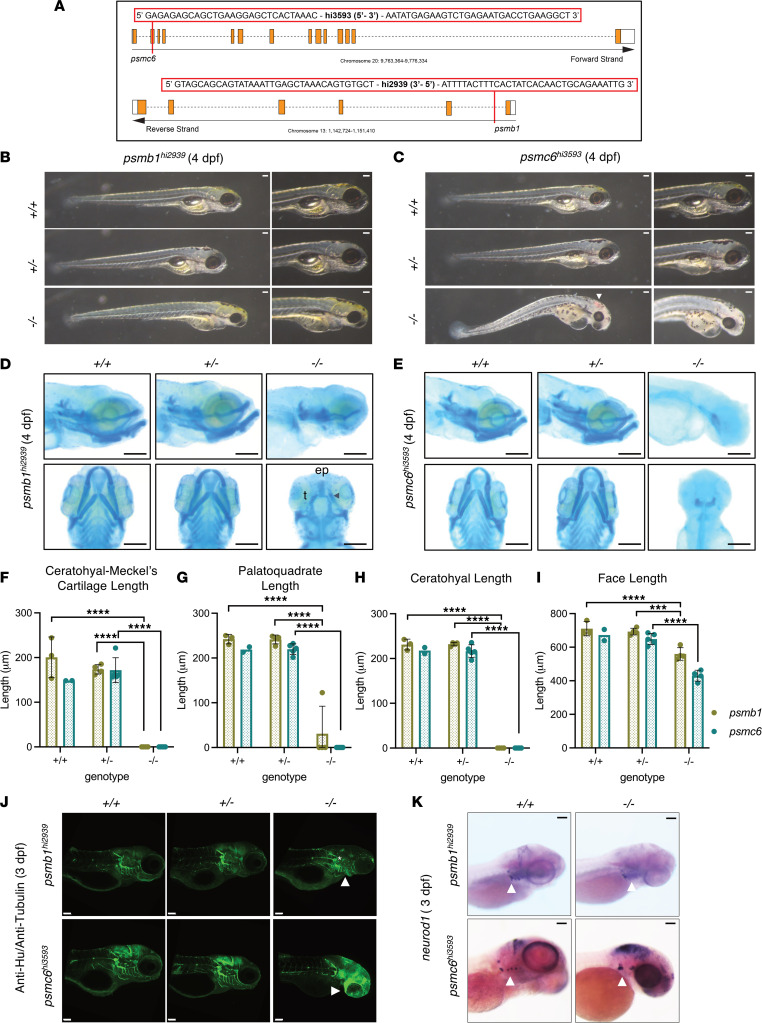
Figure 4
(A) Map of the transgenic insertion sites for psmb1hi2939 and psmc6hi3593 mutants. (B and C) Widefield imaging of psmb1hi2939 and psmc6hi3593 mutants. Both have craniofacial malformations, and psmc6–/– larvae have cardiac edema and blood pooling in the brain (C, white arrowhead). (D and E) Widefield imaging of Alcian blue–stained larvae. (D) psmb1–/– larvae (4 dpf) lack cartilages contributing to the lower jaw and have a reduced ethmoid plate (ep) and abnormal trabecula (t). Cartilage remnants (arrowhead) from the lower jaw appear in a subset of homozygotes. (E) psmc6–/– lack most cranial cartilage. (F–I) Measurements of craniofacial features obtained from ImageJ analysis of Alcian blue–stained larvae (4 dpf; ***P ≤ 0.001, ****P ≤ 0.0001, ordinary 1-way ANOVA with Dunnett’s multiple-comparison test following an observation of a difference in measurements between +/+ and –/– embryos; psmb1+/+, n = 3; psmb1+/–, n = 4; psmb1–/–, n = 4; psmc6+/+, n = 2; psmc6+/–, n = 5; and psmc6–/–, n = 5). (J) Anti-Hu/Anti-tubulin IHC at 72 hpf. psmb1–/– and psmc6–/– have abnormal brain structure and cranial ganglia (white star) and nerve development (white arrowheads). (K) ISH for neurod1 at 72 hpf. psmb1–/– and psmc6–/– have abnormal and missing vagal/cranial ganglia (white arrowheads). Scale bars: 100 μm. Data shown represent mean ± SD.