
|
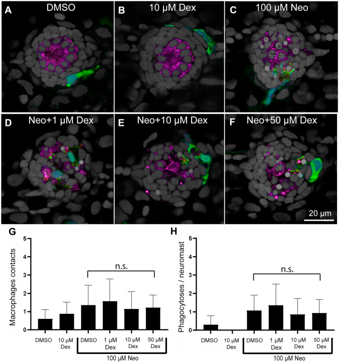
Fig. 5
Treatment with dexamethasone did not inhibit macrophage response to neuromast injury. A–F Representative confocal images of control (A, B) and neomycin injured neuromasts (C–F). Larvae (6 dpf) were treated for 24 h with 1, 10, or 50 µM dexamethasone, then exposed for 30 min to 100 µM neomycin to induce rapid neuromast injury. After drug exposures, all specimens were fixed and processed for labeling of macrophages (green, YFP), hair cells (magenta, Otoferlin) and nuclei (gray, DAPI). Normal localization of macrophages was observed in control fish (A, B) and no inhibition was observed in neuromasts treated with neomycin (C–F). G, H Dexamethasone pretreatment did not affect macrophage contacts with dying hair cells (G, P > 0.9999, Dunn’s multiple comparisons test) or the number of phagocytotic events (H, P > 0.9999, Dunn’s multiple comparisons test). Bars indicate SD. N = 15 fish/group; 2 experimental trials