
|
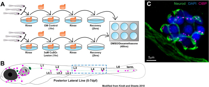
Fig. 1
Workflow of CuSO4 lateral-line lesion and dexamethasone exposure. A Diagram of workflow for CuSO4 lesions and dexamethasone exposure. Larvae (5 dpf) were placed into cell strainers and moved between rinse and lesion steps, while being allowed to swim freely during recovery and dexamethasone exposure. B Diagram of a 5–7 dpf zebrafish larvae showing the distribution of lateral-line neuromasts (magenta) and innervating afferent nerves (green). This study focused on neuromasts L3, L4, and L5 (blue dashed-line box). C Representative maximum intensity projection of confocal z-stack image of neuromast L4 in a 7 dpf zebrafish larvae. Labels: afferent neurons labeled with GFP (green; tgBAC (neurod1: GFP), DAPI-labeled hair cell nuclei (blue), and immunolabeled presynaptic ribbons (magenta; pan-CtBP)