Figure 3
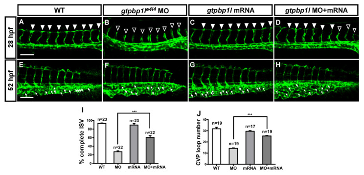
Overexpression of gtpbp1l rescues the loss of gtpbp1l. (A) In wt control embryos, ISV grows toward the dorsal and formed DLAV at approximately 28–30 hpf (arrowheads). However, ISVs showed slowed or stalled growth at mid-somite in gtpbp1le4i4 MO ((B), hollow arrowheads). (C) Overexpression of gtpbp1l by low-dosage gtpbp1l mRNA injection (0.012 ng) showed no defects in vasculature; however, overexpressed gtpbp1l can rescue the growth defect of ISV ((D), solid arrowheads). (E–H) At 52 hpf, fewer endothelial cells sprouted, and loop formation occurred at the CVP in gtpbp1l MO (white arrows in (F)) compared to the wt control (E). Injection of gtpbp1l mRNA induced no obvious defect in CVP (G) but rescued the defect of CVP loops (H). (I) Quantification of the percentage of embryos with completed ISV at 28 hpf shows an increase of ~32% in rescued embryos compared to gtpbp1l morphants. The percentages of embryos with completed ISV were ~93 ± 7 (n = 23), 28 ± 8 (n = 22), 90 ± 12 (n = 23), and 60 ± 16 (n = 22) in the control, gtpbp1l MO knockdown, gtpbp1l mRNA overexpression, and rescued embryos, respectively. (J) Quantification data of CVP formation were ~27 ± 3 (n = 19), 12 ± 3 (n = 19), 26 ± 4 (n = 17), and 17 ± 3 (n = 19) in the control, gtpbp1l MO knockdown, gtpbp1l mRNA overexpression, and rescued embryos, respectively. *** indicates p < 0.0001 by unpaired Student’s t-test. Data are mean ± S.D. Scale bar = 100 μm for (A–H).

