
Fig. 6
- ID
- ZDB-IMAGE-221222-13
- Publication
- Chen et al., 2021 - Acute brain vascular regeneration occurs via lymphatic transdifferentiation
- All Figures
- Figures for Chen et al., 2021
|
Fig. 6
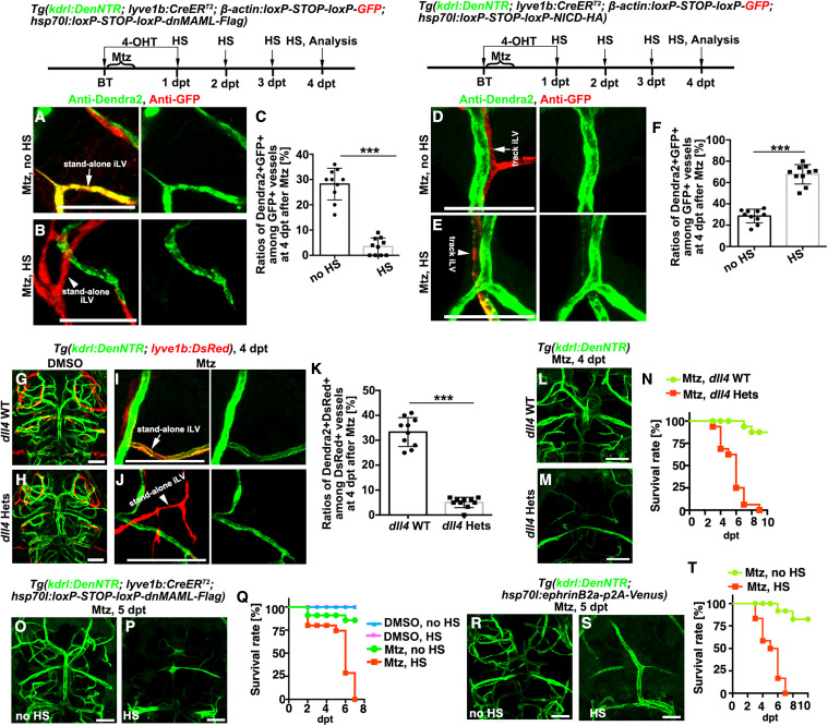
Figure 6. The stand-alone iLV-to-BV transdifferentiation is dependent on Notch and critical for postinjury survival (A–C) Transdifferentiation of the stand-alone iLVs (A, arrow) was inhibited by the LEC-specific overexpression of dnMAML-Flag (B, arrowhead). The statistics show the ratios of Dendra2+GFP+ vessels among all the GFP+ vessels (C) (n = 10 larvae; two-tailed unpaired t test; ∗∗∗, p < 0.0001). (D–F) The track iLVs (D, arrows) were induced to undergo LV-to-BV transdifferentiation by the LEC-specific overexpression of NICD-HA (E, arrowhead). The statistics show the ratios of Dendra2+GFP+ vessels among all the GFP+ vessels (F) (n = 10 larvae; two-tailed unpaired t test; ∗∗∗, p < 0.0001). (G–K) In contrast to the wild-type (G), the uninjured dll4 heterozygotes (H) were physiologically normal including brain BVs and BLECs. But after Mtz treatment, the stand-alone iLV-to-BV transdifferentiation (I, arrow) failed to occur in the dll4 heterozygotes (J, arrowhead). The statistics show ratios of Dendra2+GFP+ vessels among all the GFP+ vessels (K) (n = 10 larvae; two-tailed unpaired t test; ∗∗∗, p < 0.0001). (L–T) The dll4 heterozygotic mutation (L–N), the LEC-specific overexpression of dnMAML-Flag (O–Q), and general overexpression of EphrinB2a (R–T), led to defects in the formation of early-regenerated BVs and became lethal. The statistics show the survival rates (N, n = 32; Q, n = 40; T, n = 36). Note that the majority of the larvae with defective formation of early-regenerated BVs died before 6 dpt, while all of them died by 7 dpt. Scale bar, 50 μm. Data are represented as mean ± SD. Hets, heterozygotes; HS, heat shock. See also Figures S6 and S7.
Reprinted from Developmental Cell, 56(22), Chen, J., Li, X., Ni, R., Chen, Q., Yang, Q., He, J., Luo, L., Acute brain vascular regeneration occurs via lymphatic transdifferentiation, 3115-3127.e6, Copyright (2021) with permission from Elsevier. Full text @ Dev. Cell