Fig. 6
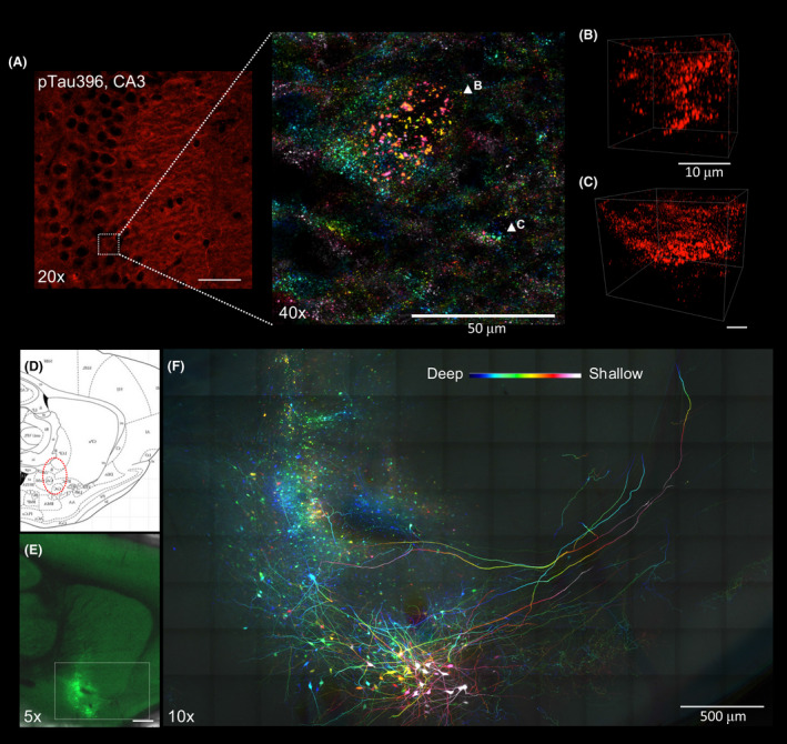
Applications of Accu‐OptiClearing in rodent brain tissues. Visualisation of phosphorylated tau aggregation in 3xTg mice. (A) Image of the hippocampal CA3 region stained with anti‐phosphorylated tau antibody. Inset showing colour‐coded projection of phosphorylated tau‐positive aggregations (indicated by arrowheads in B and C). Scale bar, 50 μm (for A), 10 μm (for B and C). Long‐range tracing of rat nucleus basalis of Meynert neurons with AAV‐GFP. (D) Sagittal rat brain atlas indicating the nbM region in red dotted circle. (E) Tiled Z‐stack image showing AAV‐GFP‐labelled nbM neurons three weeks after injection into the nbM region (scale bar = 1 mm). (F)Colour‐coded projection image with higher magnification of the region shown in E (scale bar = 500 μm, z‐depth = 205 μm)

