
Fig. 2
- ID
- ZDB-IMAGE-221221-2
- Publication
- Glasauer et al., 2021 - DNA template strand segregation in developing zebrafish
- All Figures
- Figures for Glasauer et al., 2021
|
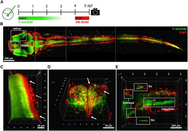
Fig. 2 Figure 2. Whole-mount analysis of label retaining and proliferating body regions using light sheet microscopy (A) Experimental approach where zygotes were injected with 5.7 pmol F-ara-EdU, followed by soaking in 10 mM BrdU from 4-5 dpf, fixation and staining. Maximum intensity light sheet projections of a whole zebrafish: (B) dorsal view of whole animal composed of four merged images, (C) anterior view of eye, (D) anterior view of forebrain, and (E) dorsal view of the mid-body region. See Supplemental Video 1 showing 3-D representation. Arrows indicate examples of BrdU positive / Fara-EdU negative cells.