
|
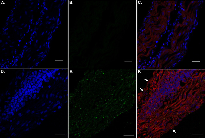
Fig 7 The presence of nApoE41-151 within tail regions of zebrafish embryos.
A-C: Representative images from confocal immunofluorescence in 5 mm paraffin-embedded sections of non-treated control 48 hpf zebrafish embryos that were stained with DAPI (A), anti-His antibody (1:500) (B), and the merged image together with PHF-1 (1:250) in Panel (C). There was no detection of any nApoE41-151 fragments in untreated control sections as indicated by the lack of labeling in Panel B. D-F: Identical to Panels A-D with the exception that embryos were exogenously treated for 24 hours with 25 μg/ml of nApoE41-151. In this case punctate staining of the fragment was observed that appeared cytoplasmic. PHF-1 labeling was identified in muscle cells that exhibited abnormal morphology (arrows, Panel F). All scale bars represent 20 μm.