
|
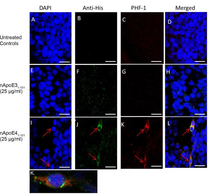
Fig 5 Tau pathology present after treatment with exogenous nApoE41-151 fragment in 72 hpf zebrafish brain.
Representative 40X images from confocal immunofluorescence in 5 mm paraffin embedded sections of non-treated control 72 hpf zebrafish (A-D), nApoE31-151-treated at 25 μg/ml (E-H), or nApoE41-151-treated at 25 μg/ml (I-L). Strong PHF-1 labeling was only observed following treatment with nApoE41-151 (I-L). Panel K depicts a separate, representative merged image following treatment with nApoE41-151. In this case, at high magnification the fibrillar nature of PHF-1 labeling was apparent. All scale bars represent 50 μm. Data are representative of three independent experiments.