
Fig 7
- ID
- ZDB-IMAGE-221216-28
- Publication
- Zhao et al., 2022 - Genetic analysis of activin/inhibin β subunits in zebrafish development and reproduction
- All Figures
- Figures for Zhao et al., 2022
|
Fig 7
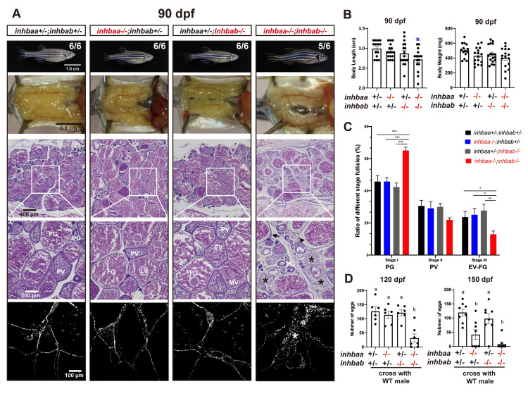
(A) Morphology, gross anatomy and histological analysis of activin βA female mutants at 90 dpf. Follicles from the control (inhbaa+/-;inhbab+/-) and single mutants (inhbaa-/-, inhbab-/-) showed normal growth and ovarian development. The double mutant (inhbaa-/-;inhbab-/-) started to show stromal cell hyperplasia (asterisk), granulosa cell hypertrophy (arrow) and follicle atresia (arrowhead). The overproliferation of the stromal cells is also shown by DAPI staining for nuclei (bottom). The boxed areas are shown at higher magnification below. The numbers shown in the photos indicate the total number of fish examined (lower) and the fish exhibiting similar phenotype to that shown (upper). (B) Body length and body weight of activin βA mutants at 90 dpf. (C) Follicle composition in the ovaries of the activin βA mutants at 90 dpf (n = 5). (*p < 0.05; **p < 0.01; ***p < 0.001). PG, primary growth (stage I); PV, previtellogenic (stage II); EV-FG, vitellogenic (stage III). (D) Fertility and fecundity of activin βA female mutants at 120 and 150 dpf. The mutant females were bred with normal WT males by natural breeding. Different letters indicate statistical significance (p < 0.05). Each dot represents the average egg number of four females from each spawning test (n = 6-8 tests).