
Figure 3
- ID
- ZDB-IMAGE-221214-34
- Genes
- Publication
- Faustini et al., 2022 - Synapsin III Regulates Dopaminergic Neuron Development in Vertebrates
- All Figures
- Figures for Faustini et al., 2022
|
Figure 3
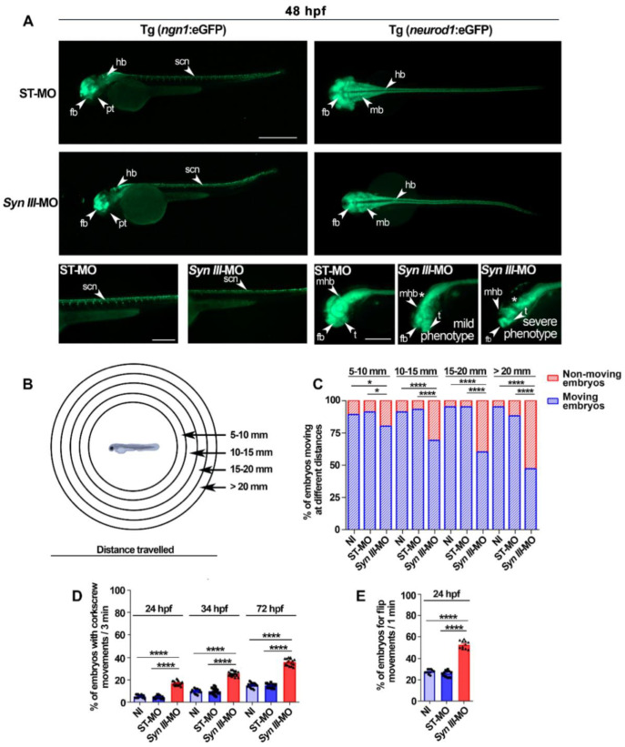
Syn III-MO injection affects Tg(ngn1:eGFP) and Tg(neurod1:eGFP) embryos neurodevelopment and behavior. (A) Representative lateral (left panels) and dorsal (right panels) views of ngn1 and neurod1 dependent eGFP fluorescence in the 48 hpf ST-MO- or Syn III-MO-injected Tg(ngn1:eGFP) and Tg(neurod1:eGFP) embryos, respectively. Full lateral/dorsal view scale bar, 500 μm. Tail/head magnifications scale bars = 250 μm. Asterisk indicates the midbrain/hindbrain regions exhibiting a clear reduction of the eGFP signal in the morphants. Abbreviations: fb, forebrain; hb, hindbrain, mb: midbrain; mhb, midbrain hindbrain boundary; t, telencephalon; pt, posterior tuberculum; scn, spinal cord neurons. (B) Schematic representation of the touch-evoked test. (C) Swimming performance of embryos at 72 hpf in the touch-evoked test. A significant progressive decrease in the % of syn3 KD morphants when compared to NI or ST-MO-injected embryos was detected (* p < 0.05, **** p < 0.0001, one-way ANOVA + Newman-Keuls, results are representative of three independent experiments with 40 embryos per group). (D) A significant increase in the percent of Syn III-MO-injected embryos exhibited corkscrew movement when compared to NI or ST-MO-injected embryos was observed at 24, 48 or 72 hpf (**** p < 0.0001, one-way ANOVA + Newman-Keuls, results are representative of three independent experiments with ≅ 10 embryos per group). (E) A significant increase in flipping movement was observed in the Syn III-MO-injected embryos when compared to NI or ST-MO-injected ones at 24 hpf (**** p < 0.0001, one-way ANOVA + Newman-Keuls, results are representative of three independent experiments with 10 embryos per group).