IMAGE

Figure 3

ID
ZDB-IMAGE-221214-221
Source
Figures for Ursu et al., 2022
Image
Figure Caption

Figure 3

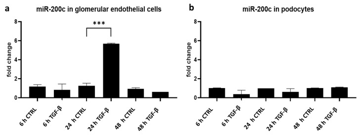
TGF-β stimulation induces miR-200c expression in cultured glomerular endothelial cells. (a,b) qPCR for miR-200c detection in cultured human glomerular endothelial cells (a) and human podocytes (b) normalized to U6 RNA. Data are given as fold change compared to unstimulated cells (CTRL) at the respective time point. Cells were stimulated with TGF-β for 6 h, 24 h and 48 h, *** p < 0.001. Reproducible data from three independent experiments.

Acknowledgments
This image is the copyrighted work of the attributed author or publisher, and ZFIN has permission only to display this image to its users. Additional permissions should be obtained from the applicable author or publisher of the image. Full text @ Int. J. Mol. Sci.