
Image
|
Figure Caption
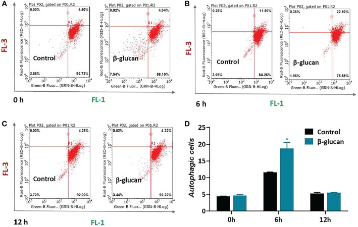
Figure 6 Effects of β-glucan on autophagy level in ZF4 cells. Autophagy was quantified by flow cytometry after AO staining. The flow cytometry results of control or β-glucan-treated ZF4 cells at 0 h (A), 6 h (B), and 12 h (C) post SVCV infection were exhibited; (D) Percentage of control or β-glucan-treated ZF4 cells positive for autophagosomes at different time points after viral infection. Values represent the means ± SEM. *P < 0.05 (n = 6).
Acknowledgments
This image is the copyrighted work of the attributed author or publisher, and
ZFIN has permission only to display this image to its users.
Additional permissions should be obtained from the applicable author or publisher of the image.
Full text @ Front Immunol