
|
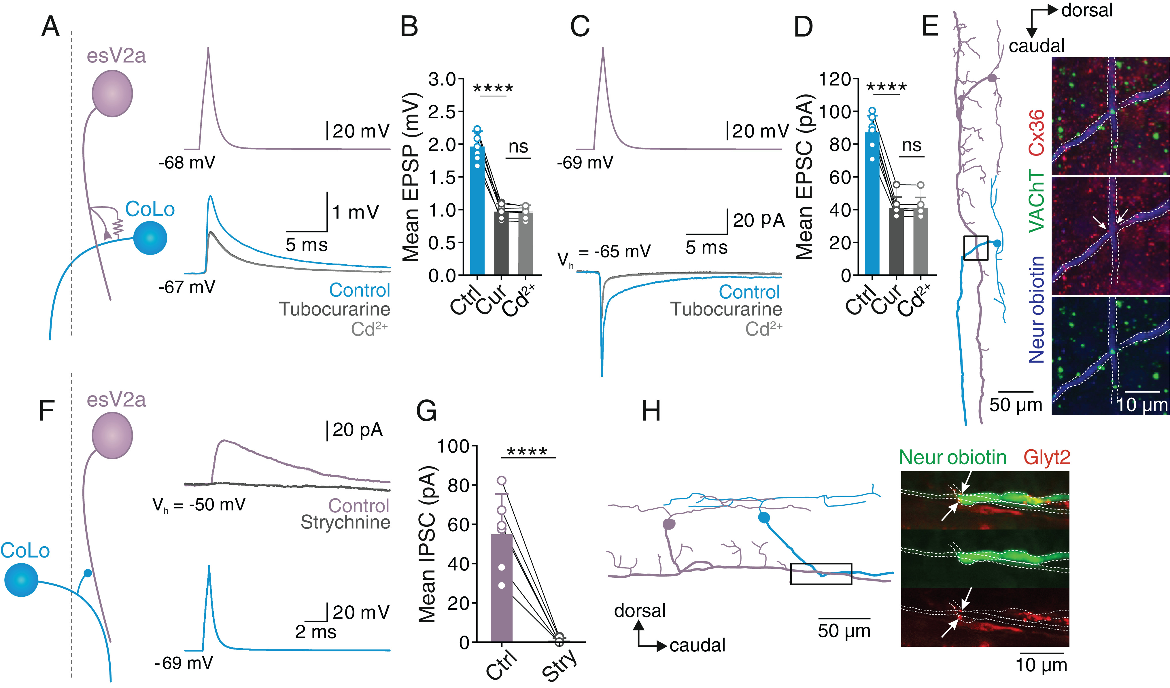
Fig. 7 Feedforward excitation and inhibition between esV2a interneurons and CoLo interneurons, respectively. (A) Dual whole-cell patch-clamp recording from a rostral esV2a interneuron and an ipsilateral caudal CoLo interneuron (the dashed line indicates the middle line of spinal cord). esV2a interneurons made mixed electrical and chemical synapses with CoLo interneurons. (B) Quantification of the mean amplitude of monosynaptic EPSPs in CoLo interneurons. The graph shows mean ± SD, n = 7; one-way ANOVA, P = 7.5 × 10⁻⁵ for control vs. d-tubocurarine; P > 0.05 for d-tubocurarine vs. Cd2+. (C) The same pair of esV2a interneuron and CoLo interneuron as in A in which EPSCs were examined. (D) Quantification of the mean amplitude of the monosynaptic EPSCs elicited in CoLo interneurons. Graph represents mean ± SD, n = 7; one-way ANOVA, P = 8.2 × 10⁻⁵ for control vs. d-tubocurarine; P > 0.05 for d-tubocurarine vs. Cd2+. Each dot represents the data from single neurons. (E) Reconstruction of a recorded esV2a interneuron and CoLo interneuron (Left). Confocal image showing the coexpression of VAChT (green) and Cx36 (red and indicated by white arrows) at the site of synaptic contacts between the axons of the two interneurons (blue and indicated by the dashed lines). These images correspond to the region indicated in the box of the Left panel. (F) Dual whole-cell patch-clamp recording of an esV2a interneuron and a contralateral CoLo interneuron (the dashed line indicates the middle line of spinal cord). Single action potentials in CoLo interneuron elicited IPSCs in esV2a interneuron that were blocked by strychnine. (G) Quantification of the effect of strychnine on the mean amplitude of monosynaptic IPSCs. Data are presented as mean ± SD, n = 6; paired t test, P = 6.3 × 10⁻⁷. Each dot represents the data from a single neuron. (H) Lateral view of the reconstruction of the pair of CoLo and esV2a interneuron shown in F. GlyT2 was expressed at the site of presumed synaptic contact between CoLo and esV2a interneuron axon (arrows).