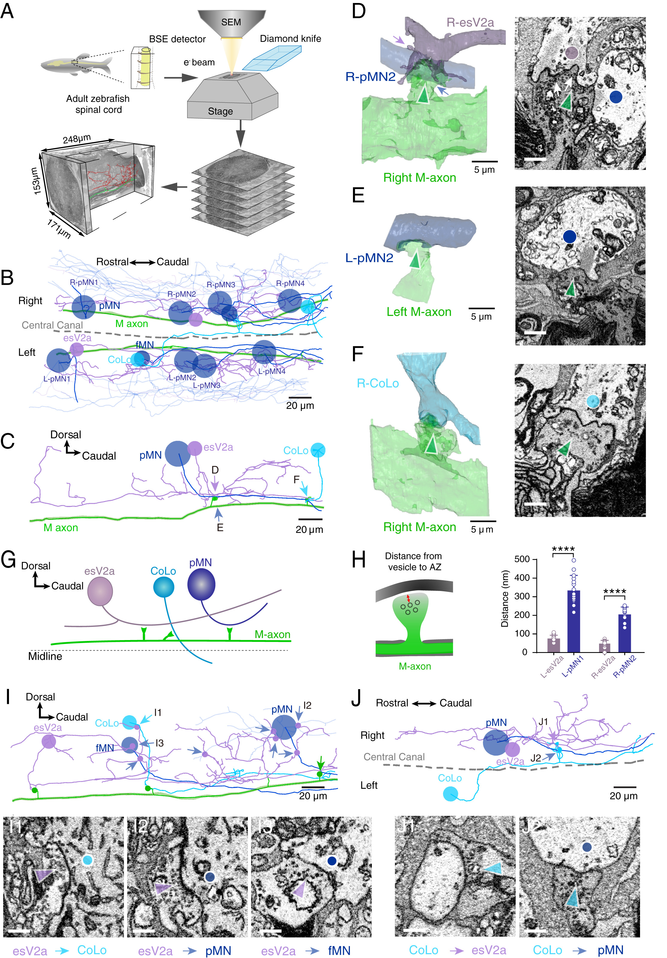
Fig. 4 Connectome of spinal escape neural circuit revealed by SBEM. (A) Workflow of acquiring three-dimensional electron microscopy volume from adult zebrafish spinal cord. The images were centered at the central canal and covered the entire spinal cord cross-section; 7,095 cuts along the axial direction were performed, resulting in a final dimension of 171 × 153 × 248 μm3. Skeletonized reconstruction was done by human annotations. (B) Skeletonized neurites of eight pMNs (navy blue, larger soma size), two esV2a interneurons (purple), two CoLo interneurons (sky blue), two fMNs (navy blue, smaller soma size), as well as two M-axons (green) contained in the dataset. Dashed line indicating the spinal central canal. (C) The contralateral view of connectomes of the right M-axon (green), R-esV2a interneuron (purple), R-pMN2 (navy blue), and R-CoLo (sky blue) in the right side of the spinal cord. The collateral of M-axon made putative axo-axonal synaptic contacts with the R-esV2a interneuron (purple), the R-pMN2 (navy blue), and the R-CoLo interneurons (sky blue). (D) Volume reconstruction (Left) and electron micrographs (Right) of the indicated synaptic contacts in C (purple and navy blue arrow). The second collateral of the right M-axon made synaptic contacts with both the R-esV2a interneuron and the R-pMN2. Vesicle cluster (indicated by white arrows) in the terminal of the M-axon collateral (Right, green arrow) was found in the vicinity of the R-esV2a interneuron (Right, purple dot), but not of the pMN2 (Right, navy blue dot). (E) Volume reconstruction (Left) and electron micrograph (Right) of the indicated synaptic contact. The third collateral of the left M-axon made synaptic contacts with axon of a L-pMN2. No vesicle cluster in the terminal of M-axon collateral (Right, green arrow) was found in the vicinity of the L-pMN1 axon (Right, navy blue dot). (F) Volume reconstruction (Left) and electron micrograph (Right) of the indicated synaptic contacts in C. The fifth collateral of the right M-axon made synaptic contacts with the R-CoLo interneuron. No vesicle cluster in the terminal of right M-axon collateral (Right, green arrow) was found in the vicinity of the R-CoLo axon (Right, sky blue dot). (G) The wiring diagram between right M-axon with the three types of spinal interneurons in the right side of the spinal cord. M-axon formed putative synapses with the esV2a interneurons, the CoLo interneuron and pMNs. (H, Left) The drawing showing the distance between vesicles and presynaptic membrane. (Right) Statistic results suggest the different distance for esV2a interneurons and pMNs (Student’s t test, n = 30; P = 1 × 10⁻⁶ for L-esV2a and L-pMN1; n = 30; P = 5 × 10⁻⁵ for R-esV2a and R-pMNs). Each dot represents the data from single vesicles. (I) Lateral view of connectomes of a presynaptic L-esV2a interneuron (purple) with a postsynaptic L-CoLo interneuron (sky blue), L-pMN4 (navy blue), and L-fMN (navy blue) on the left side of spinal cord in B. Chemical synapses (purple dots) were formed between the axon of presynaptic L-esV2a interneuron and the axon of the postsynaptic L-CoLo interneuron (sky blue), L-pMN4 (navy blue, larger soma size), and L-fMN (navy blue, smaller soma size). Electron micrographs (I1, I2, and I3) showing the indicated synaptic contacts. Colored arrows indicating the location and standing for the neuronal types. (J) Connectomes of a presynaptic L-CoLo interneuron with a postsynaptic R-esV2a interneuron (purple) and a postsynaptic R-pMN2 (navy blue) on the contralateral side of spinal cord in the dorsal view. The presynaptic L-CoLo interneuron made axo-axonic synapses (sky blue dots) with the contralateral pMN (navy blue) and cholinergic esV2a interneuron (purple). Electron micrographs (J1 and J2) showing the indicated synaptic contacts. Colored arrows indicating the location and standing for the neuronal types. (Scale bars in B, C, I, and J: 20 μm; in D–F, Left: 5 μm; D–F, Right: 500 nm; and in I1 to I3 and J1 and J2: 500 nm.)
Image
Figure Caption
Acknowledgments
This image is the copyrighted work of the attributed author or publisher, and
ZFIN has permission only to display this image to its users.
Additional permissions should be obtained from the applicable author or publisher of the image.
Full text @ Proc. Natl. Acad. Sci. USA

