
|
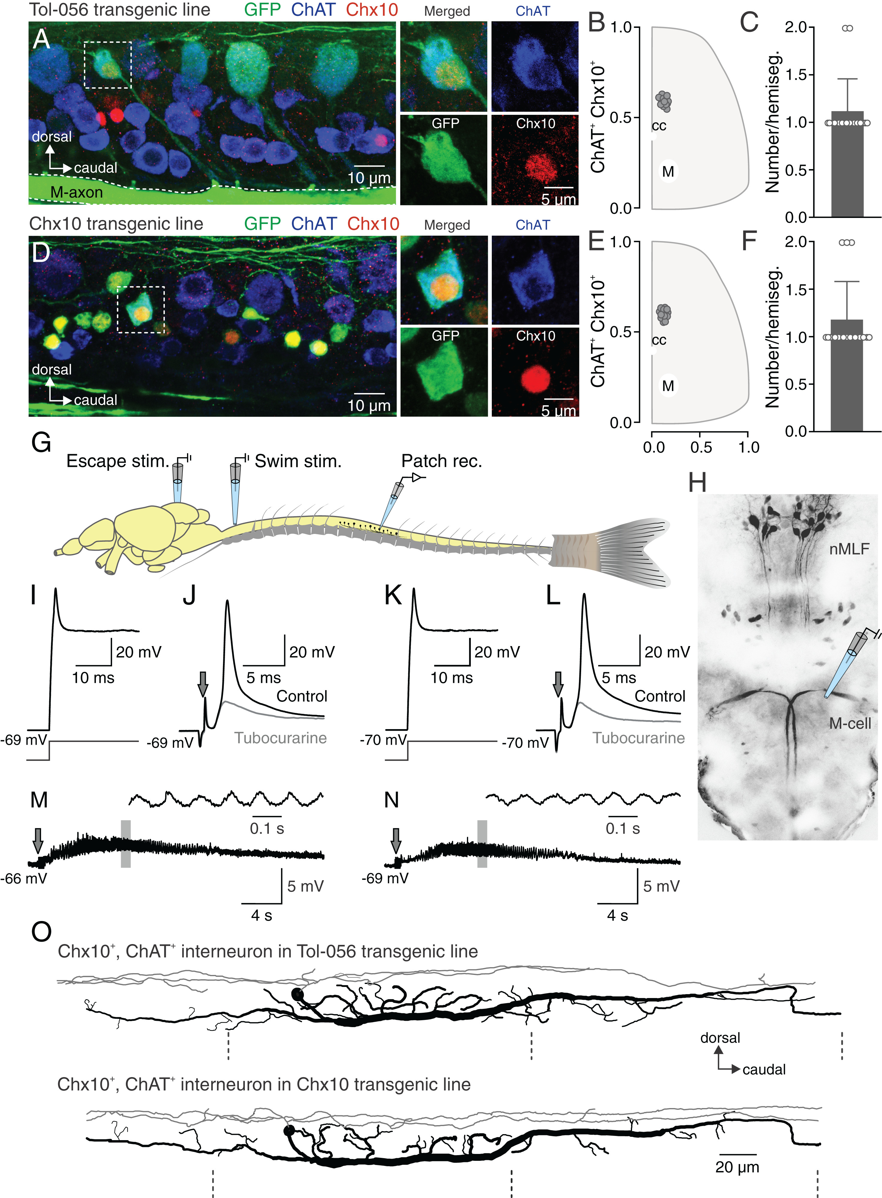
Fig. 1 esV2a interneurons involved in escape behavior. (A) Lateral view of a spinal segment from Tol-056 enhancer trap transgenic zebrafish line with immunohistochemical staining. The dashed box indicates a specific interneuron expressing GFP (green) that colocalizes both Chx10 (red) and ChAT (blue). This neuron is shown specifically on the right four boxes. (B) Distribution of esV2a interneurons in the spinal cord from the Tol-056 transgenic line (cc: central canal; M: M-axon). Data are from multiple fish and multiple segments. (C) Quantification of esV2a interneurons per hemisegment in Tol-056 transgenic line. The graph shows mean ± SD; n = 16. (D) Lateral view of a spinal segment from Tg(Chx10:GFP) transgenic line with immunohistochemical staining. A specific GFP expressing neuron (green) in the dashed box was immunoreactive for both Chx10 (red) and ChAT (blue) shown specifically in the right four boxes. Yellow represents noncholinergic V2a interneurons that express GFP (green) and Chx10 (red). (E) Distribution of esV2a interneurons in the spinal cord from Tg(Chx10:GFP) transgenic line (cc: central canal; M: M-axon). (F) Quantification of esV2a interneurons per hemisegment in Tg(Chx10:GFP), the graph shows mean ± SD; n = 16. (G) Experimental setup of the ex vivo preparation showing the sites for extracellular stimulation and whole-cell patch-clamp recordings. (H) Dorsal view of descending neurons in the zebrafish hindbrain revealed by retrograde labeling from the spinal cord. (I) esV2a neuron in Tol-056 transgenic line discharged a single action potential in response to a prolonged depolarizing current injection. (J) The same esV2a neuron as in I, receiving a strong cholinergic cEPSP from an M-cell that was blocked by d-tubocurarine leaving on an eEPSP. Arrow indicates electrical stimulation. (K) An esV2a neuron in Tg(Chx10:GFP) firing a single action potential in response to a prolonged depolarizing current injection. (L) The same esV2a neuron as in K receiving a strong cholinergic cEPSP by an M-cell stimulation that was blocked by d-tubocurarine leaving only an eEPSP. Arrow indicates electrical stimulation. (M) An esV2a interneuron recorded in the Tol-056 line was not recruited during swimming activity (same interneuron as in I and J). Arrow indicates electrical stimulation. (N) An esV2a interneuron recorded in the Tg(Chx10:GFP) line was not recruited during swimming activity (same interneuron as in K and L). Arrow indicates electrical stimulation. (O) Morphology of an esV2a neuron in Tol-056 line (Upper) and Tg(Chx10:GFP) (Lower). Gray line representing the dendrite and black representing the soma and axonal projections. The dashed lines indicating the segmental boundaries.