
Fig. 8
- ID
- ZDB-IMAGE-221127-8
- Publication
- Webster et al., 2021 - Role of BMP signaling during early development of the annelid Capitella teleta
- All Figures
- Figures for Webster et al., 2021
|
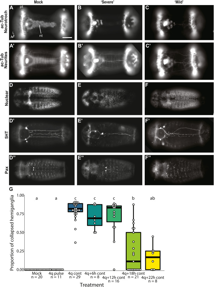
Fig. 8 Fig. 8. Trunk neural phenotype in cont BMP-treated (Z-stack). (A,D) A mock animals showing wild-type phenotype. (B,E) 4q+22 h continuous animals showing a severe VNC collapse phenotype. (C,F) 4q+22 h continuous animals showing a mild VNC collapse. (A–C) acetylated-tubulin showing neurotroch cilia, (A′-C′) acetylated-tubulin in the VNC. (D–F) Hoechst nuclear stain. (D′-F′) 5HT (D″-F″) Pax, arrowhead: normally bilaterally-symmetric cells on either side of the midline in each segment. Figure panels with the same letter represent the same animal. A, anterior, L, left side, nt, neurotroch; pt, prototroch; tt, telotroch; VNC, ventral nerve cord; scale bar = 50 μm (G) Boxplot showing the significant effect of treatment on proportion of segments with collapsed hemiganglia, letters indicate significance groups.
Reprinted from Developmental Biology, 478, Webster, N.B., Corbet, M., Sur, A., Meyer, N.P., Role of BMP signaling during early development of the annelid Capitella teleta, 183-204, Copyright (2021) with permission from Elsevier. Full text @ Dev. Biol.