
|
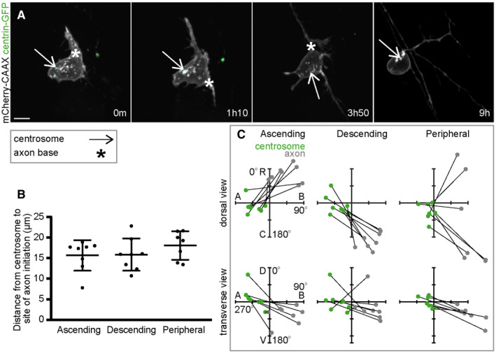
Figure EV2
A. Image sequence from confocal time lapse shows a Rohon‐Beard neuron labelled with membrane and centrosome markers during the initiation of the ascending (0 m), descending (1 h10 min) and peripheral axons (3 h 50 min), and during axon pathfinding. The centrosome is located away from the base of each axon but moves close to the peripheral axon during pathfinding. Images are maximum projections from confocal z‐stacks. Scale bar = 10 μm. B. Graph showing distance between centrosome and base of the axon at time of initiation of each axon ( C. Plots showing the positions of the centrosome and base of the axon at the time of axon initiation relative to the cell centroid at 0,0 for dorsal and transverse views for ascending (