
Fig. 3
- ID
- ZDB-IMAGE-221115-12
- Genes
- Publication
- Lawir et al., 2019 - Evolutionary transition from degenerate to nonredundant cytokine signaling networks supporting intrathymic T cell development
- All Figures
- Figures for Lawir et al., 2019
|
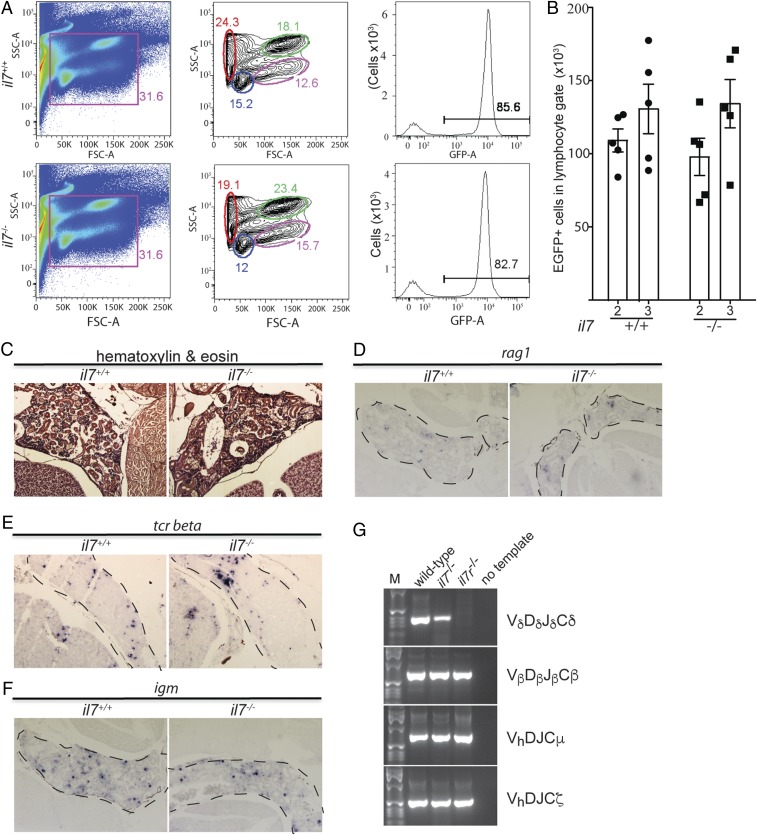
Fig. 3
Characterization of hematopoiesis in 3-mo-old zebrafish kidneys. (A) Flow cytometry analysis of cell populations in whole kidney marrow of adult wild-type and il7-mutant fish (Left and Middle). The different cell populations as identified by the characteristic light-scatter characteristics are indicated (red, erythrocytes; blue, lymphocytes; green, myeloid cells; pink, precursor cells) (34). The majority of cells in the lymphocyte gate (blue) are GFP-positive (Right). (B) Enumeration of GFP-positive cells in the lymphocyte gate of wild-type and il7-mutant fish at age 2 and 3 mo. Each data point represents 1 fish; data are mean ± SEM. (C) Normal histology of kidney marrow in il7 mutant adult fish. (D–F) RNA in situ hybridization on tissue sections of adult wild-type and il7 mutant kidney marrow (outlined with dashed lines) with probes specific for rag1 (D), indicative of B cell precursors; tcr beta (E), indicative of αβ T cells; and igm (F), indicative of Β cells. (G) RT-PCR assay for expressed completely assembled antigen receptor genes. Vd-Dd-Jd-Cd assemblies associated with the γδ T cell lineage are present in the whole kidney marrow of il7 mutants but not in that of il7r mutants; assemblies of other lineage-specific antigen receptors are present in both mutant fish lines (Vb-Db-Jb-Cb for αβ T cells, Vh-D-J-Cm for Igμ-expressing B cells, and Vh-D-J-Cz for Igζ-expressing B cells). Representative panels are shown for wild-type fish (n = 5), il7 mutants (n = 4), and il7r mutants (n = 5).