
Fig. 6
- ID
- ZDB-IMAGE-221108-6
- Publication
- Kowatschew et al., 2021 - An ancient adenosine receptor gains olfactory function in bony vertebrates
- All Figures
- Figures for Kowatschew et al., 2021
|
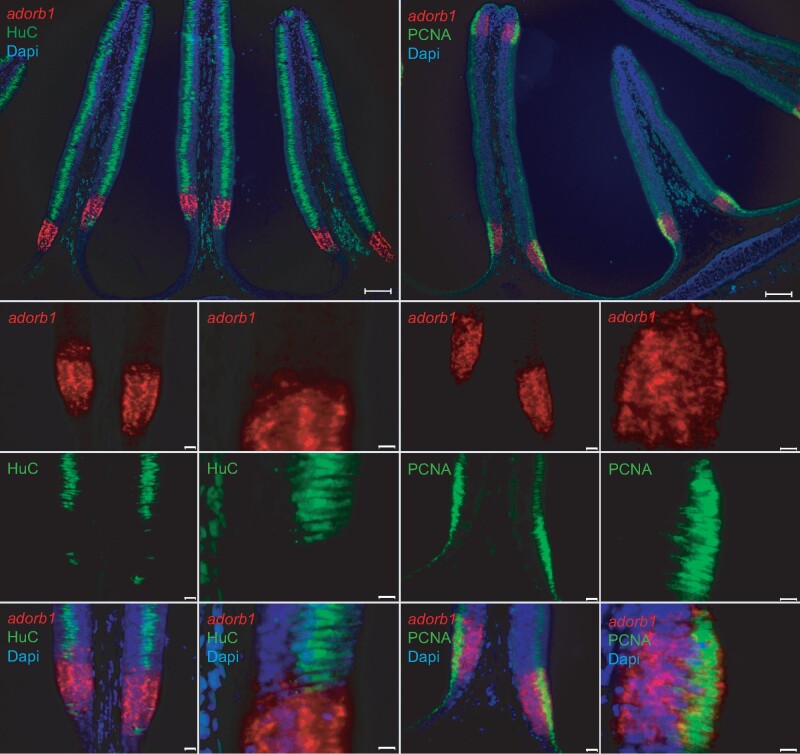
Fig. 6
Lamprey adorb1 expression zone borders the sensory area of the olfactory organ. Double labeling of horizontal cross-sections of L. fluviatilis olfactory epithelium with HuC antibody and PCNA antibody confirms the expression of adorb1 (visualized by ISH) in a nonsensory region. Genes and color as indicated on the respective panels, nuclei are visualized by DAPI staining (blue). The neuronal marker HuC (left columns, green) labels the olfactory sensory area of the lamella, whereas PCNA (right columns, green) labels the adjacent mitotic regions. Adorb1 (red) is expressed in a contiguous manner in the PCNA-positive region, but absent from the sensory region. Note that PCNA labeling (green) is restricted to the apical layer, whereas adorb1(red) staining is distributed more broadly. Top panels show an overview (scale bars 100 µm); the smaller bottom panels are enlarged (scale bars 50 and 10 µm, respectively).