
|
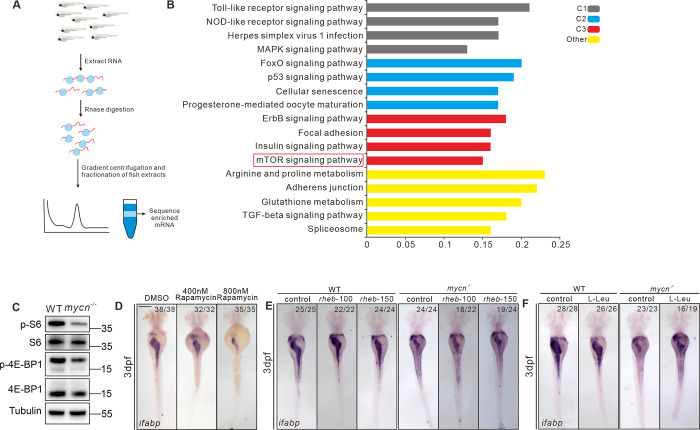
Fig 7
(A) Schematic representation of the experimental workflow of the Ribo-seq. (B) KEGG analysis of the genes showing reduced translation efficiency in the mycn mutant. The red box marks the mTOR pathway. (C) Detection of the rps6, phosphorylated rps6, eIF4EBP, and phosphorylated eIF4EBP in WT and mycn mutant embryos at 3 dpf. The proteins were detected by western blot. The tubulin expression level was used as internal control. (D) Inhibiting the mTOR pathway by different doses of rapamycin mimicked the intestinal defects phenotype of the mycn mutants. (E, F) Activating the mTOR pathway by injecting rheb mRNA and L-Leu treatment partially rescued the intestinal defects phenotype of the mycn mutants. All embryos are shown in dorsal view. Scale bar: 50 μm (C), 200 μm (D-F). Raw images of this figure are provided in S1 Raw Images. dpf, days postfertilization; ifabp, intestine fatty acid–binding protein; WT, wild-type.