
|
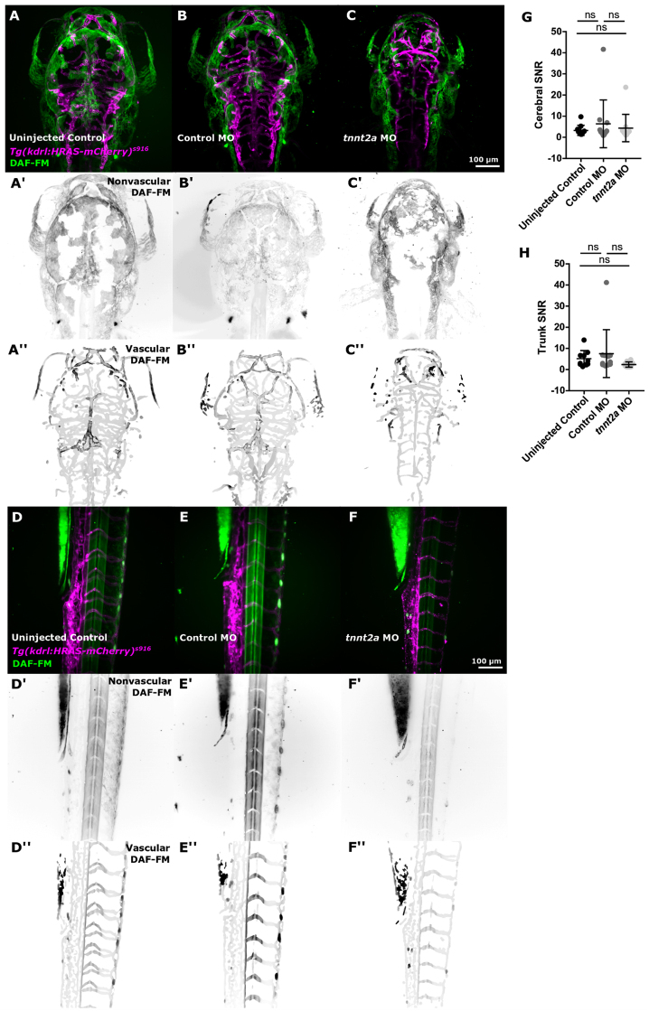
Fig. 8
Inflammation is not increased by absent blood flow. (A, B and C) Examining nitric oxide (NO; visualized by DAF-FM) as an inflammatory marker, showed similar levels of NO in the head of uninjected controls, control MO, and tnnt2a MO (uninjected control n = 11, control MO n = 12, tnnt2a MO n = 11; three experimental repeats; 3dpf). (D, E and F) DAF-FM levels in the trunk vasculature appeared visually similar between groups. (G) Quantification of cerebral DAF-FM SNR showed no difference between tnnt2a MO and uninjected controls (P > 0.9999) or control MO (P > 0.9999; Kruskal–Wallis test). (H) Quantification of trunk DAF-FM SNR showed no difference between tnnt2a MO and uninjected controls (P = 0.1391) or control MO (P = 0.1216; Kruskal–Wallis test).