
|
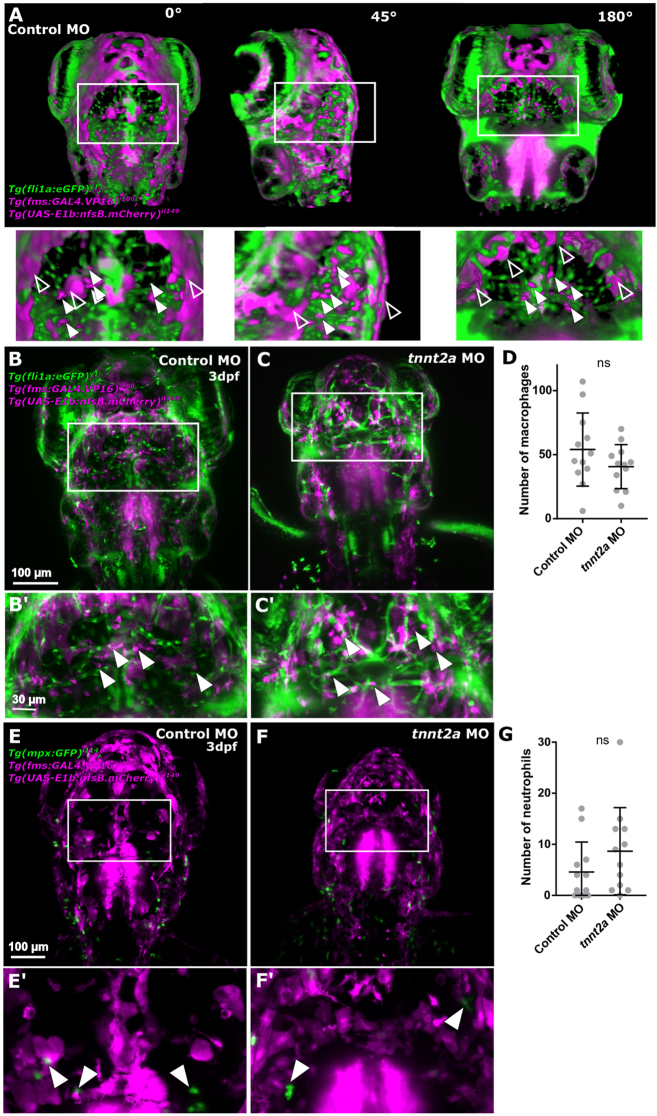
Fig. 7
Lack of blood flow does not impact the number of cerebral innate immune cells. (A) Identification of immune cells was performed in 3D, allowing to discern non-specific signal (unfilled arrowhead) from immune cells (filled arrowhead). (B and C) Macrophages (magenta) were quantified examining the transgenic Tg(fli1a:eGFP)y1, Tg(fms:GAL4.VP16)i186, Tg(UAS-E1b:nfsb.mCherry)il149. (D) Number of macrophages was not changed upon blood flow loss (P = 0.2356; n = 12; 3dpf; Mann–Whitney U test). (E and F) Neutrophils (green) were examined in the transgenic reporter line Tg(mpx:GFP)i114, Tg(fms:GAL4.VP16)i186, Tg(UAS-E1b:nfsb.mCherry)il149. (G) Number of neutrophils was not changed upon blood flow loss (P = 0.1708; n = 12; two experimental repeats; 3dpf; Mann–Whitney U test).