
|
Figure 1
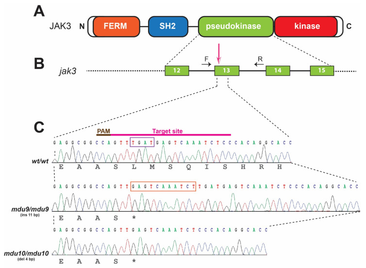
Generation of zebrafish carrying Jak3 mutations resembling those found in autosomal-recessive SCID. (A). Schematic representation of the JAK3 protein showing FERM (orange), SH2 (blue), pseudokinase (green) and kinase (red) domains. (B). Intron-exon structure of the zebrafish jak3 gene region encoding the pseudokinase domain, with the area targeted denoted with the pink arrow and the genotyping primers indicated by black arrows (F: forward, R: reverse). Exons are shown as numbered boxes and introns as solid lines. (C). Nucleotide sequence of zebrafish homozygous for wild-type (wt) and mutant (mdu9 and mdu10) alleles of jak3, with their protein translations displayed below in black text and the CRISPR/Cas9 target site shown above. The mdu9 allele represents an 11 bp insertion (orange box) and the mdu10 allele a 4 bp deletion (purple box), both resulting in frameshifts that introduce a stop codon at the equivalent location within the pseudokinase domain.