Fig. 5
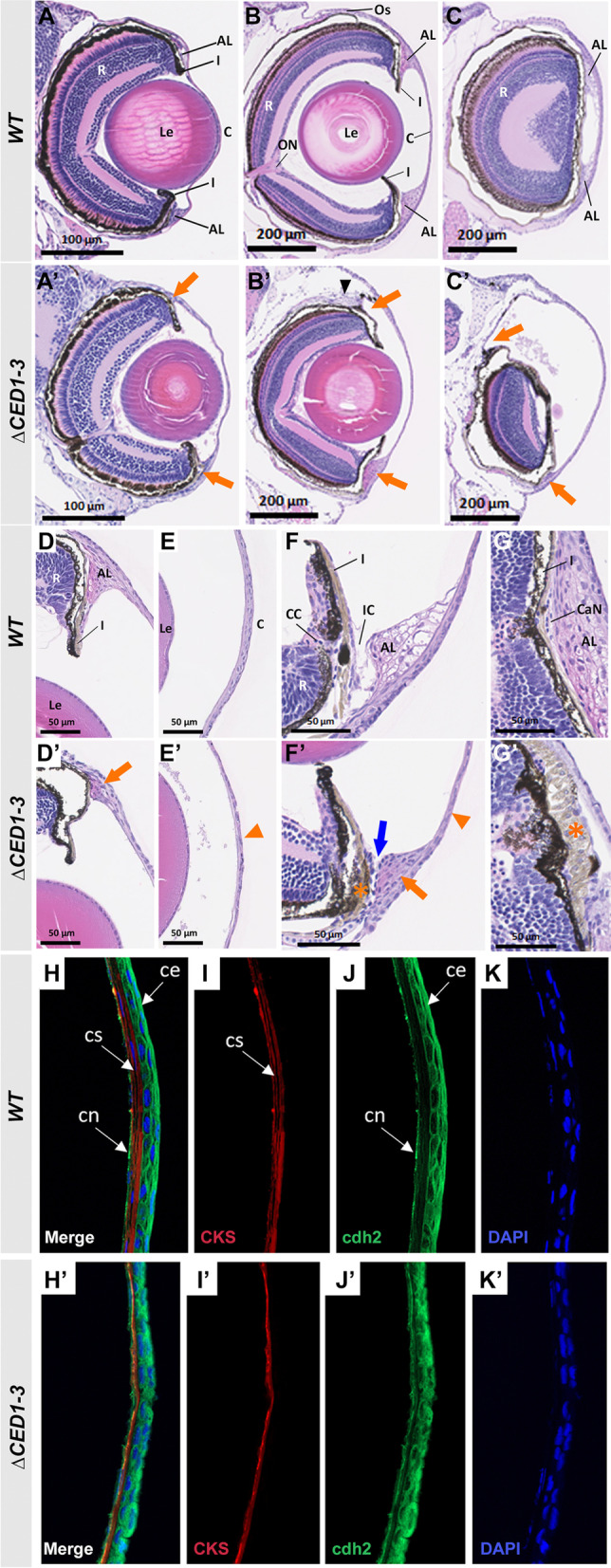
Histological analysis of ocular anomalies in foxc1a∆CED1−3 homozygous embryos. A, A’ H&E-stained transverse sections of the eye of 6-dpf wild-type and mutant embryos. B–C’ H&E-stained transverse sections through central (B and B’) and nasal (C and C’) eye regions of 30-dpf wild-type and mutant fish. Mutants show a marked enlargement of the anterior chamber and abnormal development of both dorsal and ventral annular ligaments (orange arrows, A’–C’); dislocation of lenses toward the back of the eye (B’); and deformed/misplaced scleral ossicles at the dorsal irido-corneal angle (black arrowhead, B’). D–E’ 20× magnifications of the dorsal irido-corneal angle (D and D’) and cornea (E and E’) showing details of the hypoplastic dorsal annular ligament (orange arrow, D’), and thin cornea at 30-dpf (orange arrowhead, E’). Transverse (F and F’) and coronal (G and G’) 40× magnifications of the ventral irido-corneal angle and canalicular network showing an apparent absence of the glycoprotein aggregates in the ventral annular ligament (orange arrow in F’), narrowing of the irido-corneal canal (blue arrow, F’), hyperplasia of the ventral iris stroma in this region (orange asterisks in F’ and G’) and thin cornea at 30-dpf (orange arrowhead in F’). H–K’ immunostaining of cornea sections of 30-dpf wild-type and mutant fish with anti-CKS (red) and anti-cdh2 (green), showing a thinner corneal stroma (I’) and a disorganized corneal epithelium (J’). AL, annular ligament; C, cornea; CaN, canalicular network; CC, ciliary canal; ce, corneal epithelium; cn, corneal endothelium; cs, corneal stroma; I, iris; IC, irido-corneal canal; Le, lens; ON, optic nerve; Os, scleral ossicle R, retina

