
Image
|
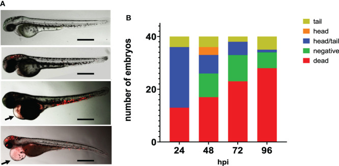
Figure Caption
Figure 2
Fluorescence microscopy of zebrafish embryos at 28 hpf infected with mCherry expressing H44/76_B meningococci (5000 cfu).
Acknowledgments
This image is the copyrighted work of the attributed author or publisher, and
ZFIN has permission only to display this image to its users.
Additional permissions should be obtained from the applicable author or publisher of the image.
Full text @ Front Cell Infect Microbiol