Fig. 5
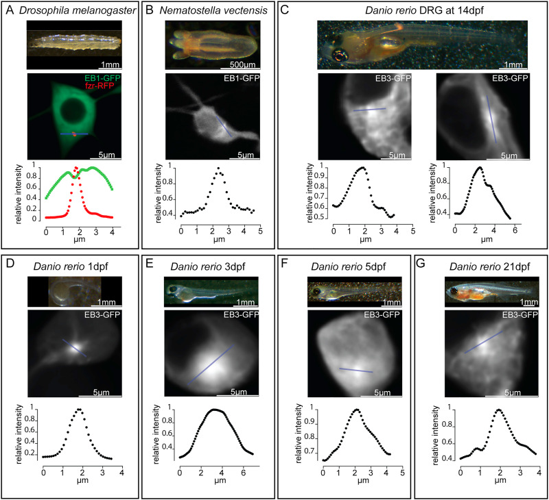
Fig. 5. Zebrafish sensory neurons maintain a somatic hotspot of microtubule organization. A. Brightfield image of a 3rd instar drosophila larva and image of a ddaE neuron cell body expressing EB1-GFP and fzr-RFP are shown. The blue line indicates the region used to quantify EB1-GFP intensity from a sum projection of a timeseries. The output for fzr-RFP (red) and EB1-GFP (green) is shown in the graph. B. A brightfield image of a 4–6 tentacle stage Nematostella vectensis polyp and an image of a tripolar ganglion cell expressing EB1-GFP are shown. The blue line was used as the region for summing intensity through the time course and the output is shown in the graph. C. A brightfield image of a 14dpf zebrafish and images of DRG neuron cell bodies expressing EB3-GFP are shown together with lines used to quantify EB3-GFP fluorescence. The graphs show intensity along the line summed through the timecourse. D.-G. Brightfield overviews and images of RB neuron cell bodies expressing EB3-GFP are shown from different age fish. In each a line was used to generate summed intensity graphs.
Reprinted from Developmental Biology, 478, Shorey, M., Rao, K., Stone, M.C., Mattie, F.J., Sagasti, A., Rolls, M.M., Microtubule organization of vertebrate sensory neurons in vivo, 1-12, Copyright (2021) with permission from Elsevier. Full text @ Dev. Biol.

