
Figure 5
- ID
- ZDB-IMAGE-220930-6
- Publication
- Zhao et al., 2022 - PLAAT1 inhibits type I interferon response via degradation of IRF3 and IRF7 in Zebrafish
- All Figures
- Figures for Zhao et al., 2022
|
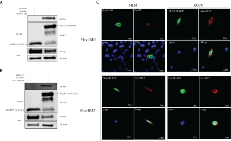
Figure 5
PLAAT1 interacts with IRF3 and IRF7. (A, B) EPC cells were transfected with the indicated plasmids (5 μg each). After 24 h, cell lysates were immunoprecipitated (IP) with α-GFP affinity resin. The immunoprecipitates and cell lysates were analyzed by immunoblotting (IB). (C) PLAAT1 co-localized with IRF3 and IRF7 in the cytoplasm. with or without SVCV infection. EPC cells were plated onto coverslips in 6-well plates and transfected with PLAAT1-GFP (2 μg) and Myc-IRF3 (2 μg) or Myc-IRF7 (2 μg) plasmids. After 24 h, the cells were left untreated (MEM), infected with SVCV. After an additional 24 h, cells were stained with DAPI (blue) and photographed under a confocal microscope. Green and red colors indicate overexpressed PLAAT1 and IRF3 or IRF7, respectively. Scale bar=25 μm. All experiments were repeated at least three times with similar results.