
Fig. 5
- ID
- ZDB-IMAGE-220913-5
- Publication
- Shrestha et al., 2022 - Embryonic Hyperglycemia Delays the Development of Retinal Synapses in a Zebrafish Model
- All Figures
- Figures for Shrestha et al., 2022
|
Fig. 5
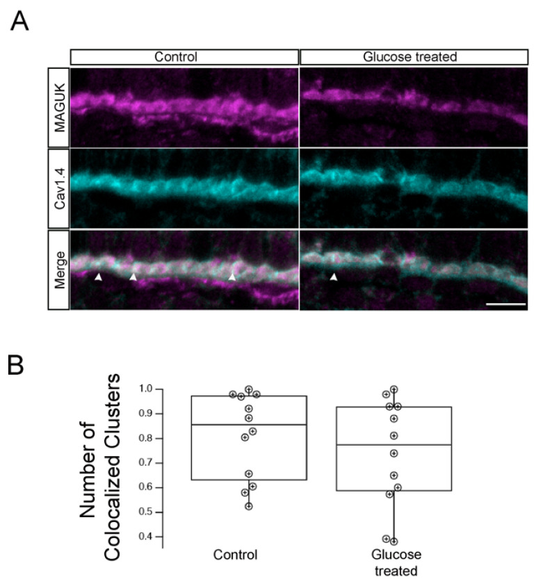
Hyperglycemic zebrafish larvae retained photoreceptor ribbon synapses with postsynaptic densities (PSDs) in INL dendrites. (A)Transverse retinal cross-sections from 5 dpf control (left panels) or hyperglycemic (right panels) larvae were double immunostained with fluorescently labeled antibodies specific for the postsynaptic marker MAGUK (magenta, top) or Cav1.4/cacna1f (cyan, middle); overlays of MAGUK and Cav1.4 labeling are shown (merge, bottom). Maximal intensity projections are shown; arrowheads indicate potential areas of colocalization. Scale bar, 5 µm. (B) Quantitative analyses of IHC for MAGUK and Cav1,4 colocalization are indicted by box-and-whisker plots. Boxes indicate interquartile ranges, while whiskers indicate range of maximal and minimal values. Scale bar, 5 µm. Abbreviations used: Cav1.4/cacna1f, voltage-dependent calcium channel; Dpf, days post-fertilization; INL, inner nuclear layer; MAGUK, membrane-associated guanylate kinase; PSD, postsynaptic densities; SEM, standard error of the mean.