
Fig. 1
- ID
- ZDB-IMAGE-220913-1
- Publication
- Shrestha et al., 2022 - Embryonic Hyperglycemia Delays the Development of Retinal Synapses in a Zebrafish Model
- All Figures
- Figures for Shrestha et al., 2022
|
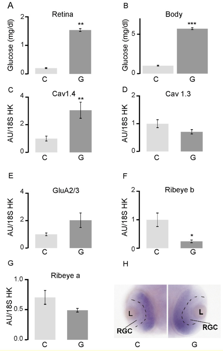
Fig. 1
In zebrafish larvae, hyperglycemia results in altered transcription of retinal synaptic genes and localization of synaptic proteins. (A) Glucose concentration at 48 hpf in the retina and (B) in the rest of the body in embryos exposed to 4% glucose 24–48 hpf; (C–G) RT-qPCR analysis of the whole retina in embryos exposed to 4% glucose 24–48 hpf. All transcript levels were normalized to those of 18S rRNA. The levels of cav1.4a transcripts increased, those of cav1.3a, GluR2/3, and ribeye a remained unchanged, while those of ribeye b decreased in retinas exposed to glucose, relative to controls; (H) In situ hybridization for ribeye a in the transverse sections in the center of the eye 48 hpf in control embryos (C) and those exposed to 4% glucose (G). The separation between the already defined retinal ganglia cell layer and the developing remainder of the retinal layer is marked by a black dashed line. * p < 0.05; ** p < 0.01; *** p < 0.001. Abbreviations: C, control; G, exposed to glucose; GluR2/3, glutamate receptor; hpf, hours post-fertilization; rRNA, ribosomal RNA; RT-qPCR, reverse transcription quantitative PCR; L, lens; RGC, retinal ganglion cells.