
Fig. 1
- ID
- ZDB-IMAGE-220912-162
- Publication
- Zhuang et al., 2022 - Histone Deacetylases Cooperate with NF-κB to Support the Immediate Migratory Response after Zebrafish Pronephros Injury
- All Figures
- Figures for Zhuang et al., 2022
|
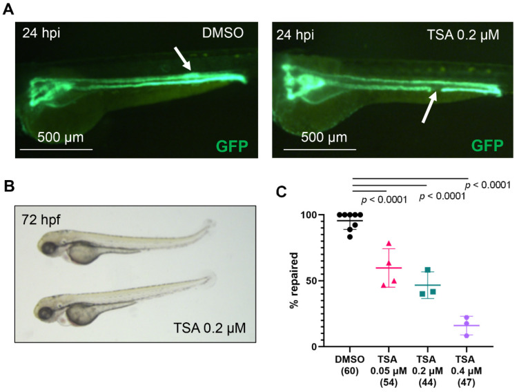
Fig. 1
Histone deacetylases are required for zebrafish pronephros repair after injury. (A) The pronephros in a control, DMSO-treated embryo of the transgenic Tg(wt1b:GFP; cdh17:GFP) zebrafish line is fully repaired at 24 h post injury (hpi, left panel). The white arrow points to the location of the repaired tubule, which is characterized by a typical thickening of the duct. In contrast, a TSA-treated embryo fails to repair the pronephros (right panel). The arrow points to the ablated portion of the tubule. (B) Zebrafish embryos, incubated in 0.2 µM TSA for one hour before ablation and 24 h post ablation developed normally. (C) TSA treatment significantly reduced the number of embryos that repaired the tubule after laser-induced injury in a concentration-dependent manner. The concentration (micromolar, µM) is shown below each group. The number of examined embryos is displayed in brackets. Individual experiments are plotted, and the mean and standard deviation for each group are displayed. Significance was calculated using Fisher’s exact test (two-tailed).