
Image
|
Figure Caption
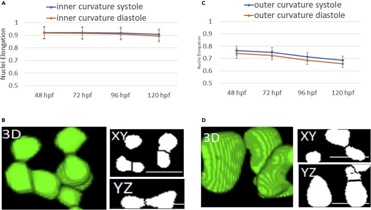
Fig. 6
Systole vs diastole circularity analysis
(A) Inner curvature nuclei are observed to have a circular shape, (symmetric circle elongation = 1, ellipse <1) with slightly higher values observed for the diastole.
(B) Outer curvature cardiomyocyte nuclei are observed to have an elongated shape with higher elongation observed in the diastole.
(C and D) Volumetric reconstructions of the circular shape of inner curvature and elliptic shape of outer curvature myocytes were visually presented, respectively. In addition, the corresponding lateral and axial views are shown with binary images (scale bar = 15 um).
Acknowledgments
This image is the copyrighted work of the attributed author or publisher, and
ZFIN has permission only to display this image to its users.
Additional permissions should be obtained from the applicable author or publisher of the image.
Full text @ iScience