
|
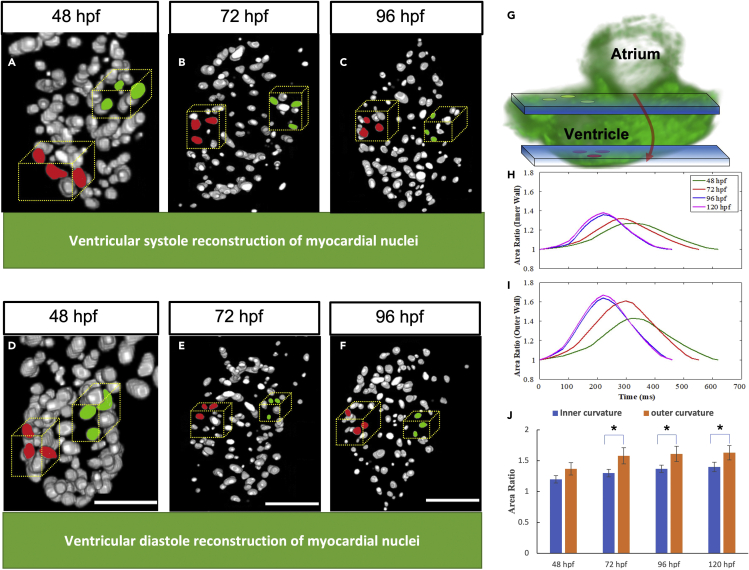
Fig. 4
Selected markers utilized area ratio analysis
(A–F) represents the systolic reconstruction of ventricular myocytes at 48 hpf, 72 hpf, and 96 hpf, respectively, while (D–F) represents the diastolic reconstruction of myocytes at different developmental stages.
(G and H) Schematic illustrating the nuclei region of interest. Blue windows represent light sheet sections. Zebrafish ventricular volumes were sampled to compare the innermost curvature contractility (green markers), with respect to the outermost curvature (red markers) (H) Area ratio for innermost curvature by tracking three cardiomyocytes highlighted green in the blue optical plane, which elucidate increasing contractility trend observed across distinct developmental stages.
(I) The area ratio for outermost curvature calculated by tracking cardiomyocytes highlighted red in the blue optical plane, indicates the outermost curvature has higher contractility compared to the innermost curvature. (J) Outermost curvature has a significantly higher area ratio compared to innermost curvature after 72 hpf (n = 3, p = 0.05, one-tail t-test).