
Image
|
Figure Caption
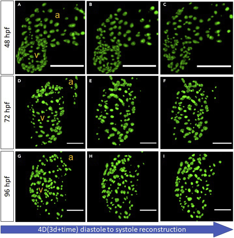
Fig. 3
Visualizing cmlc:GFPnuc zebrafish ventricular nuclei deformation at distinct developmental stages (48 – 96 h postfertilization), across the cardiac contraction cycle
(A–I) The Hessian DoG scale space representation was used for localizing cardiomyocyte nuclei ranging from different sizes, as a result of which we were able to assess ventricular contractility and complex nuclei morphology in vivo (scale bar for A-C = 100-micron, scale bar for D-I = 50 micron). A:atrium, v:ventricle.
Acknowledgments
This image is the copyrighted work of the attributed author or publisher, and
ZFIN has permission only to display this image to its users.
Additional permissions should be obtained from the applicable author or publisher of the image.
Full text @ iScience