
|
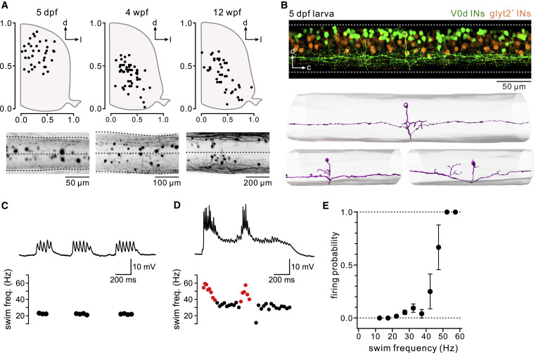
Fig. 1 Figure 1. Position, morphology, and recruitment at fast locomotor speeds of V0d interneurons in young zebrafish larvae (A) Soma positions of V0d interneurons at three different stages of development (5 days post-fertilization [dpf], 4 weeks post-fertilization [wpf], and 12 wpf) (n = 4 fish for each stage). Images below show the maximum intensity confocal projections of spinal cords from a dorsal view at each stage. (B) Top: confocal image showing V0d interneuron soma position (green) in the dorsal spinal cord in a 5-dpf zebrafish larva. Non-V0d glycinergic interneurons are shown in orange. Bottom: example morphological reconstructions of bifurcating V0d interneurons in a 5-dpf zebrafish. Asterisks mark the same neuron expressing GFP (top panel) and reconstructed (lower panel). (C) Top: patch-clamp recording of a larval (5 dpf) V0d interneuron showing a lack of recruitment at slow locomotor speeds. Bottom: a plot of instantaneous swim frequency. Black dots represent unrecruited swim cycles. (D) Top: same recording as (C) showing recruitment at fast swim speeds. Bottom: a plot of instantaneous swim frequency. Black dots represent unrecruited swim cycles, and red dots represent cycles in which the V0d interneuron was recruited. (E) Plot of firing probability against swim frequency across recorded V0d interneurons. V0d interneuron firing probability was the highest at fast locomotor frequencies (n = 12 V0d interneurons from 12 separate animals; mean and SEM). See also Figure S1.