
Fig. 6
- ID
- ZDB-IMAGE-220829-70
- Publication
- Da'as et al., 2022 - Transcriptome Profile Identifies Actin as an Essential Regulator of Cardiac Myosin Binding Protein C3 Hypertrophic Cardiomyopathy in a Zebrafish Model
- All Figures
- Figures for Da'as et al., 2022
|
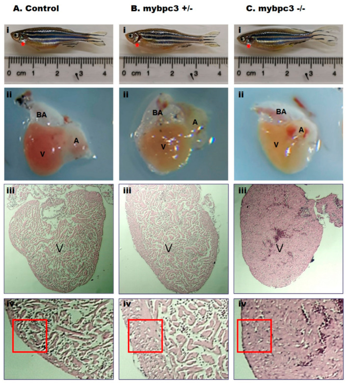
Fig. 6
The adult zebrafish mybpc3 mutant heart anatomy and histology. Adult age-matched zebrafish hearts were dissected (location: red star) and then compared across groups: (A) Control, (B) Heterozygous mybpc3+/−, and (C) Homozygous mybpc3−/− mutant. (i). Representation of the anatomical position of the heart in the adult zebrafish. (ii). The dissected heart demonstrates the two cardiac chambers, a single atrium (A), and a single ventricle (V), with an elastic, non-contractile chamber consisting of smooth muscle bulbus arteriosus (BA). (iii). Histological organization of the adult zebrafish ventricle, magnification 80×, scale bar: 10 μm. (iv). The ventricular myocardial wall is hypertrophic in the zebrafish mybpc3 mutant model (red square) when compared to the control group subset, magnification 150×, scale bar: 10 μm.