
IMAGE
Fig. 2
- ID
- ZDB-IMAGE-220829-66
- Publication
- Da'as et al., 2022 - Transcriptome Profile Identifies Actin as an Essential Regulator of Cardiac Myosin Binding Protein C3 Hypertrophic Cardiomyopathy in a Zebrafish Model
- All Figures
- Figures for Da'as et al., 2022
Image
|
Figure Caption
Fig. 2
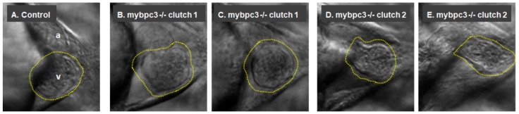
The mutant mybpc3 displayed distinct cardiac phenotypes in the zebrafish model. Representative zebrafish heart images at the ventral view at 72 hpf. (A) The control showing the cardiac chambers (atrium (a) and ventricle (v)). (B–E) The zebrafish mybpc3 mutant displayed distinct cardiac phenotypes. The zebrafish larvae from different clutches were mounted into 3% methylcellulose, and video recordings were taken using a Zeiss Axio-Zoom V16 stereomicroscope equipped with an Image Source Camera (60 frames per second) at 100× magnification, scale bar: 50 μm. A yellow traced line marks the zebrafish ventricle.
Figure Data
Acknowledgments
This image is the copyrighted work of the attributed author or publisher, and
ZFIN has permission only to display this image to its users.
Additional permissions should be obtained from the applicable author or publisher of the image.
Full text @ Int. J. Mol. Sci.