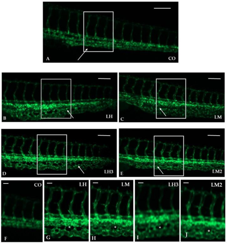
IMAGE

Figure 3

ID
ZDB-IMAGE-220828-3
Source
Figures for Zizioli et al., 2022
Image
Figure Caption

Figure 3

Purified extracts from H. aspersa caused pro-angiogenetic effects on ISVs and intercapillary spaces in the CVP: (AE) Representative images of zebrafish trunk ISVs of controls (fish water and 0.1% DMSO) (A) and treatments with 10 µg/mL of LH (B), LM (C), LH3 (D), and LM2 (E). All figures are lateral views with dorsal to the top and anterior to the left, and fluorescent vessels of embryos were observed at 48 hpf. White arrowheads indicate the CVP, showing an increase in the angiogenesis process. Enlarged white boxes indicate the magnification of ISVs and CVPs (FJ). Asterisks (*) indicate the intercapillary spaces. The experiments were repeated twice. Images were taken with a Zeiss LSM 510 META confocal laser scanning microscope (Carl Zeiss, Jena, Germany), using an objective Achroplan 10× and 20×/0.25 and a 488 nm laser, and represent 1 embryo out of 30 with the same phenotype (for more details, see Section 2).

Acknowledgments
This image is the copyrighted work of the attributed author or publisher, and ZFIN has permission only to display this image to its users. Additional permissions should be obtained from the applicable author or publisher of the image. Full text @ Curr. Iss. Mol. Biol.