IMAGE

Figure 2.

ID
ZDB-IMAGE-220814-3
Source
Figures for Zhang et al., 2022
Image
Figure Caption

Figure 2.

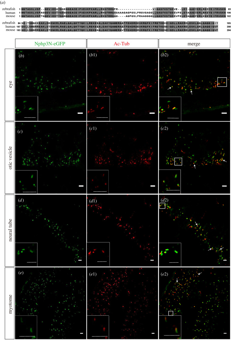
Transient expressed N-terminal peptide of zNphp3 (zNphp3N) fused eGFP localized to PC in zebrafish embryos. (a) Sequence alignment of zNphp3N with human and mouse homologs. The Clustal Omega program was used to align protein sequences from D. rerio (zebrafish), H. sapiens (human) and M. musculus (mouse). High and low amino acid similarities are emphasized in dark and light gray, respectively. (be2) At 18 hpf embryos, the transiently expressed fluorescent fusion protein zNphp3N-eGFP was precisely localized in the primary cilia of various cells in the otic vesicle, neural tube, and myotome. (b–e) Ciliary localization of zNphp3N-eGFP protein (in green) expressed transiently in zebrafish embryos. (b1–e1) Cilia labelled by anti-AcTub (red). (b2–e2) Co-location of transiently expressed zNphp3N-eGFP and cilia labelled by anti-AcTub. Arrows indicate places with obvious co-localization. The frame denotes the enlarged site. Scale bar, 5 µm.

Acknowledgments
This image is the copyrighted work of the attributed author or publisher, and ZFIN has permission only to display this image to its users. Additional permissions should be obtained from the applicable author or publisher of the image. Full text @ Open Biol.