IMAGE

Figure 5.

ID
ZDB-IMAGE-220814-1
Source
Figures for Zhang et al., 2022
Image
Figure Caption

Figure 5.

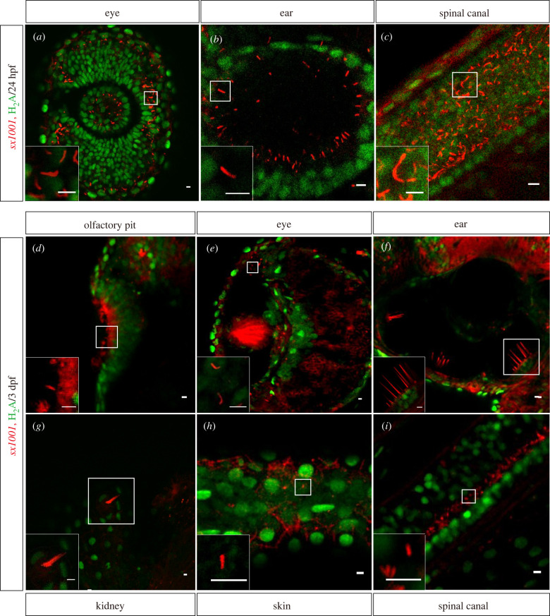
sx1001 enabled the live imaging of cilia at the tissue level. (a–c) In living sx1001 at 24 hpf, the cilia of the embryo were clearly visible in red fluorescence in the retina, otic vesicle, and spinal canal. Histone2A with C terminal fused eGFP was used to label the nucleoli in live embryos. In live 3 dpf sx1001 larvae, the various ciliary structures have been revealed and identified with a red fluorescent signal from different tissues as indicated (d–i). The frame denotes the enlarged site. Scale bar, 5 µm.

Acknowledgments
This image is the copyrighted work of the attributed author or publisher, and ZFIN has permission only to display this image to its users. Additional permissions should be obtained from the applicable author or publisher of the image. Full text @ Open Biol.