
|
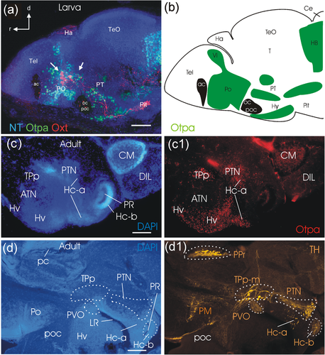
Fig. 1 Sagittal overviews of the larval zebrafish brain (5 dpf; a,b) and the adult zebrafish basal diencephalon and hypothalamus (c-d). (a) is adapted from Herget et al. (2014) and highlights oxytocin cells (white arrows pointing to red cells) in the preoptic region among Otpa (green) immunopositive cells. (b) Drawing of this brain section shows additional Otpa domains (indicated in green, brain commissures in black), for example, in the intermediate nucleus of the ventral telencephalic area (Vi, interpreted as medial amygdala; adapted from Biechl et al., 2017), in the posterior tuberculum (PT), hypothalamus(Hy), and hindbrain (HB). (c/c1) shows a parasagittal adult section close to the midline stained for DAPI (c) and Otpa (c1) identifying some posterior tubercular and hypothalamic Otpa domains. (d/d1) shows a slightly more lateral parasagittal section (d; DAPI stained) highlighting posterior tubercular and hypothalamic catecholaminergic domains (d1; TH immunostained). Scale bars = 50 µm (a), 250 µm (c/d). Abbreviations: ac, anterior commissure; ATN, anterior tuberal nucleus; Ce, cerebellum; CM, corpus mamillare; d, dorsal; DIL, diffuse nucleus of hypothalamic inferior lobe; Ha, habenula; Hc-a, caudal zone of periventricular hypothalamus in front of a posterior recess; Hc-b, caudal zone of periventricular hypothalamus around posterior recess; Hv, ventral zone of periventricular hypothalamus; Hy, hypothalamus; LH, lateral hypothalamic nucleus; LR, lateral hypothalamic recess; NT, NeuroTrace; oc, optic chiasma; Oxt, oxytocin; pc, posterior commissure; Pit, pituitary; PM, magnocellular preoptic nucleus; Po (PO in panel A), preoptic region; poc, postoptic commissure; PPr, periventricular pretectum, PR, posterior hypothalamic recess; PT, posterior tuberculum; PTN, posterior tuberal nucleus; PVO, paraventricular organ; r, rostral; T, midbrain tegmentum; Tel, telencephalon; TeO, optic tectum; TPp, periventricular nucleus of posterior tuberculum; TPp-m, magnocellular cells of TPp; TPp-p, parvocellular cells of TPp; Vi, intermediate nucleus of ventral telencephalon