
FIGURE 5
- ID
- ZDB-IMAGE-220730-38
- Publication
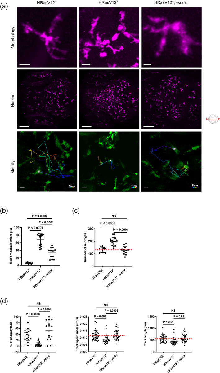
- Mazzolini et al., 2022 - Wasl is crucial to maintain microglial core activities during glioblastoma initiation stages
- All Figures
- Figures for Mazzolini et al., 2022
|
FIGURE 5