
Fig. 3
- ID
- ZDB-IMAGE-220729-3
- Publication
- Xu et al., 2021 - A neuronal circuit that generates the temporal motor sequence for the defensive response in zebrafish larvae
- All Figures
- Figures for Xu et al., 2021
|
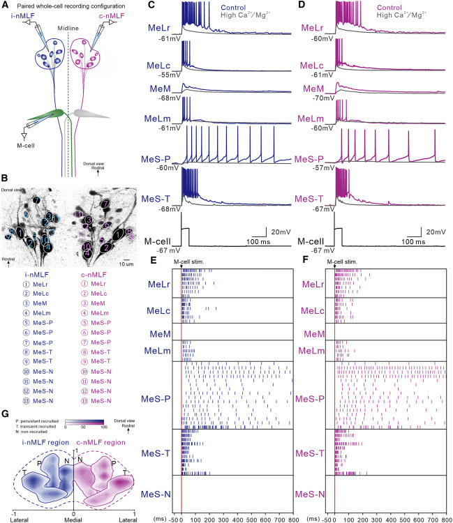
Fig. 3 Neurons in nMLF are driven by M-cell activation via indirect synaptic connection and can be divided into three functional and spatial subgroups based on their recruitment patterns (A) Line drawing illustrating the paired whole-cell, patch-clamp recording in M-cell and neurons in bilateral nMLF. (B) Image of retrograde labeling of neurons in bilateral nMLF. The numbers in the image indicate the identity of neurons in nMLF. (C and D) A single action potential generated in the M-cell induced transient discharge of action potentials in MeLr, MeLc, MeLm, as well as in MeS-T neurons and sustained discharge of action potentials in MeS-P neurons but only EPSPs in MeM neurons in both ipsilateral (blue traces in C) and contralateral (purple traces in D) nMLF. Application of high Ca2+/Mg2+ solution (gray traces in C and D) prevented or greatly reduced the firing of these neurons. (E and F) Raster plots of the transient firing pattern of action potentials for MeLr (n = 7 neurons in 7 fish), MeLc (n = 6 neurons in 6 fish), MeLm (n = 5 neurons in 5 fish), as well as MeS-T (n = 11 neurons in 11 fish) neurons; the sustained discharge of action potentials in MeS-P (n = 16 neurons in 11 fish) neurons; and lack of action potentials in MeM and MeS-N neurons in both ipsilateral and contralateral nMLF. Red vertical line indicates the time of stimulation of M-cells. (G) Contour plot of the nMLF neuron density based on different recruitment patterns by M-cell stimulation showing the spatial distribution of three subgroups (numbers of neurons for P, T, and N subgroups were 34, 42, and 47 for left nMLF and 36, 43, and 52 in right nMLF). See also Figure S1 and Videos S1 and S2.